38 gram negative cell wall diagram
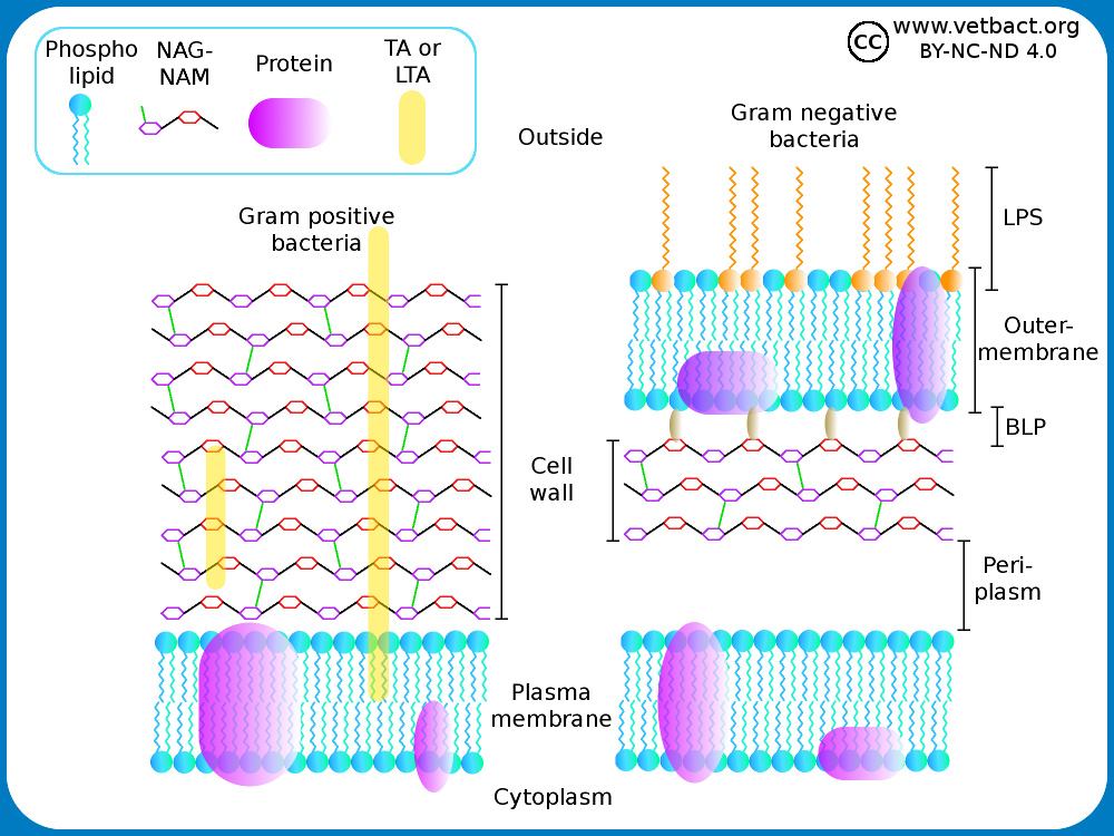
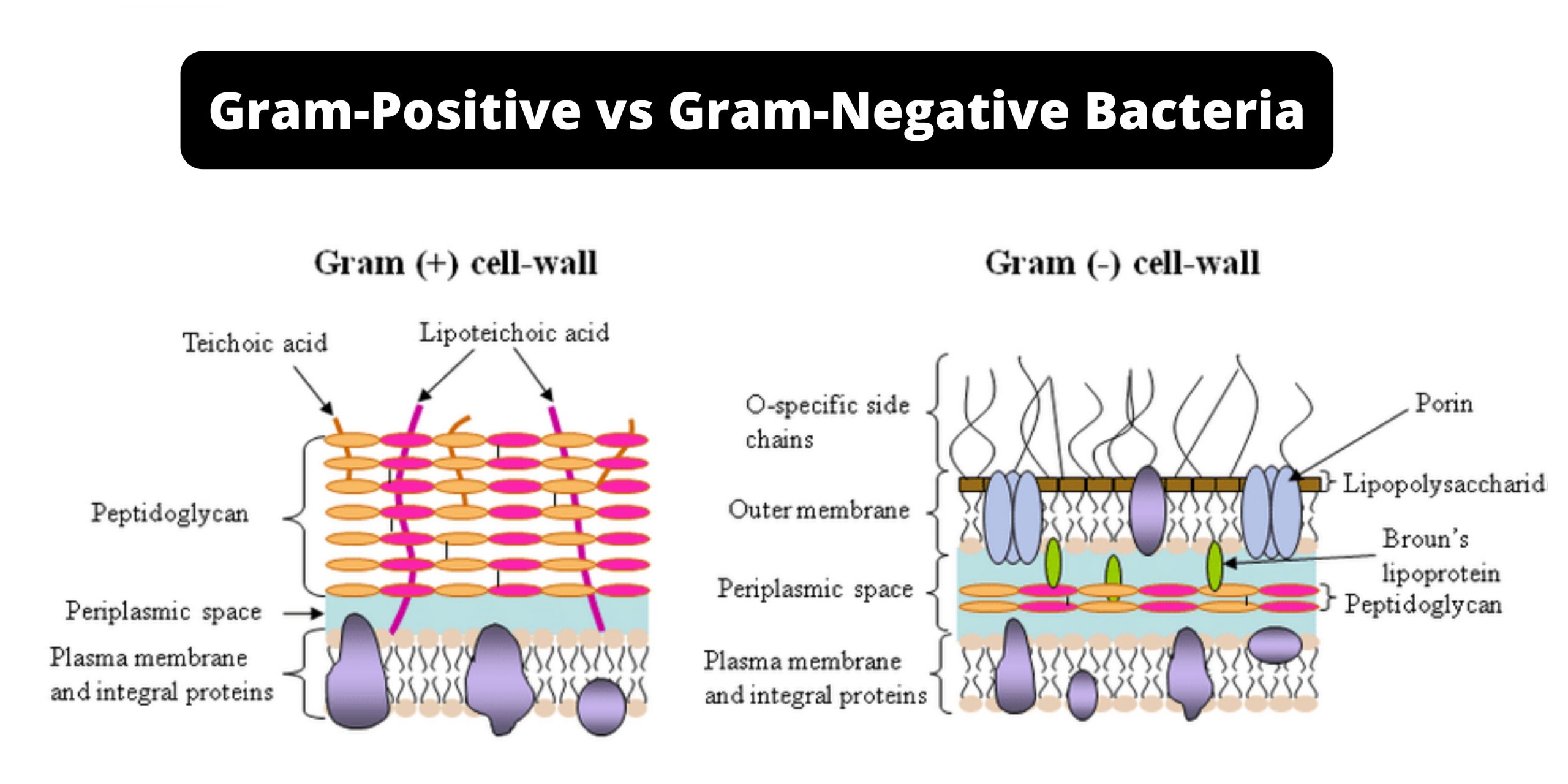
It is important to note that not all bacteria have a cell wall. Having said that though, it is also important to note that most bacteria (about 90%) have a cell wall and they typically have one of two types: a gram positive cell wall or a gram negative cell wall. From the peptidoglycan inwards all bacterial cells are very similar. Going further out, the bacterial world divides into two major classes: Gram positive (Gram +) and Gram negative (Gram -). The cell wall provides important ligands for adherence and receptor sites for viruses or antibiotics.
August 21, 2019 - One such useful classification – if a bacterium is Gram positive or Gram negative - is based on the structure of bacterial cell walls. Difference in structure of Gram positive vs Gram negative bacteria · The diagram below illustrates the differences in the structure of Gram positive and ...

Gram negative cell wall diagram
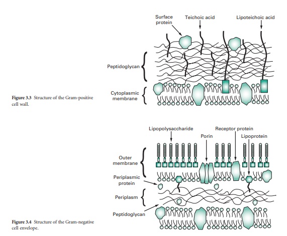
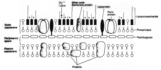
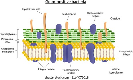
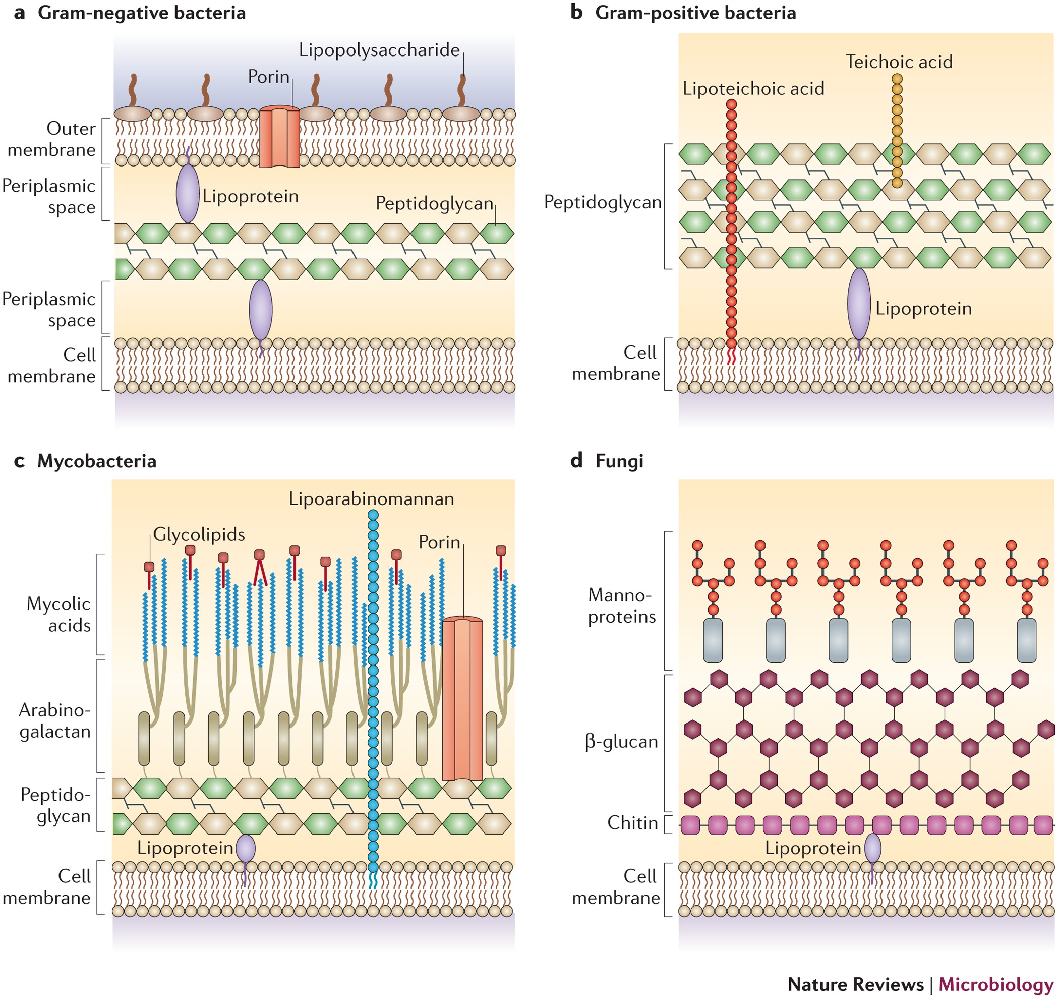
August 10, 2021 - Gram-positive cell wall. The Gram-positive cell wall is thick (15–80 nm) and more homogenous than that of the thin (2 nm) Gram-negative cell wall. The Gram-positive cell wall contains large amount of peptidoglycan present in several layers that constitutes about 40–80% of dry weight of ... Gram-negative bacteria are surrounded by a thin peptidoglycan cell wall, which itself is surrounded by an outer membrane containing lipopolysaccharide. Gram-positive bacteria lack an outer membrane but are surrounded by layers of peptidoglycan many times thicker than is found in the Gram-negatives. Gram-negative cell walls are strong enough to withstand ∼3 atm of turgor pressure (40), tough enough to endure extreme temperatures and pHs (e.g., Thiobacillus ferrooxidans grows at a pH of ≈1.5) and elastic enough to be capable of expanding several times their normal surface area (41).
Gram negative cell wall diagram. I just started using Nozin to decolonize my nose from Klebsiella aerogenes. It’s a product used in hospitals to decolonize the nose to reduce staphylococcal infections but I figured it might work for klebsiella too as gram negative bacteria are more susceptible to disinfectant types of treatment. Klebsiella and other bacteria form biofilms which protect them from traditional antibiotics, you can kill the ones outside the films and suppress the infection but the ones in the biofilm remain safe ... A mixture consisting of 100-μl cell suspension, 10 μl 0.5 M ACC solution, and 100 μl of 0.1 M Tris-HCl buffer (pH 8.5) was prepared. The mixture was incubated at 30 °C for 30 min. Then, 1 ml of 0.56 N HCl was added to the reaction mixture and centrifuged at 14,000 rpm for 5 min. 500 μl of supernatant was mixed with a mixture of 400 μl ... March 1, 2021 - It is important to note that not all bacteria have a cell wall. Having said that though, it is also important to note that most bacteria (about 90%) have a cell wall and they typically have one of two types: a gram positive cell wall or a gram negative cell wall. Gram-negative bacteria are bacteria that do not retain the crystal violet stain used in the Gram staining method of bacterial differentiation. They are characterized by their cell envelopes, which are composed of a thin peptidoglycan cell wall sandwiched between an inner cytoplasmic cell membrane ...
(I am aware that evolution doesn't work by always taking the "better" option, and that said options are only beneficial in a specific circumstance, but considering we have these two large groups then surely there must be some benefit to having the gram positive cell wall, otherwise they would be outcompeted) July 26, 2021 - Gram-negative bacteria. Gram-negative bacteria cell wall, structure, color, examples, diseases, antibiotics. Gram-negative bacteria list. January 22, 2021 - Gram staining is a technique that uses violet dye to distinguish between gram-positive and gram-negative bacteria. If the bacteria are gram-positive, the thick, peptidoglycan layer in their cell walls will retain the dye and they will stain violet. If the bacteria are gram-negative, the dye ... The sequence of steps in the Gram ... diagrammatically in Figure 2-7. Moreover, mechanical disruption of the cell wall of Gram-positive organisms or its enzymatic removal with lysozyme results in complete extraction of the CV-I complex and conversion to a Gram-negative ...
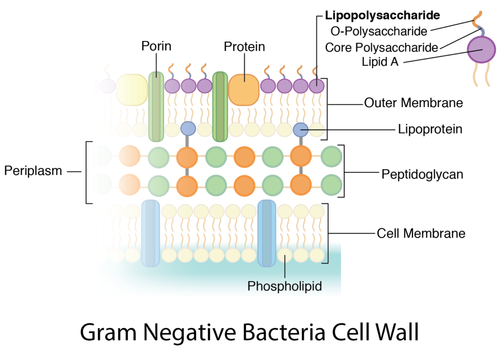
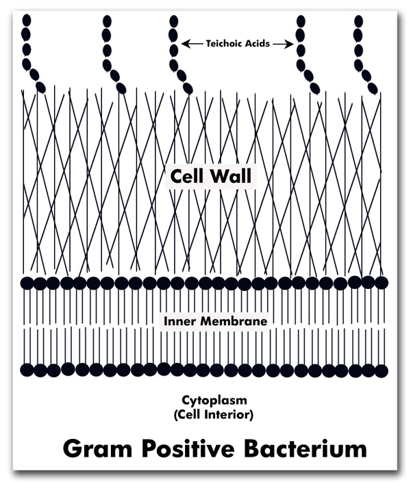
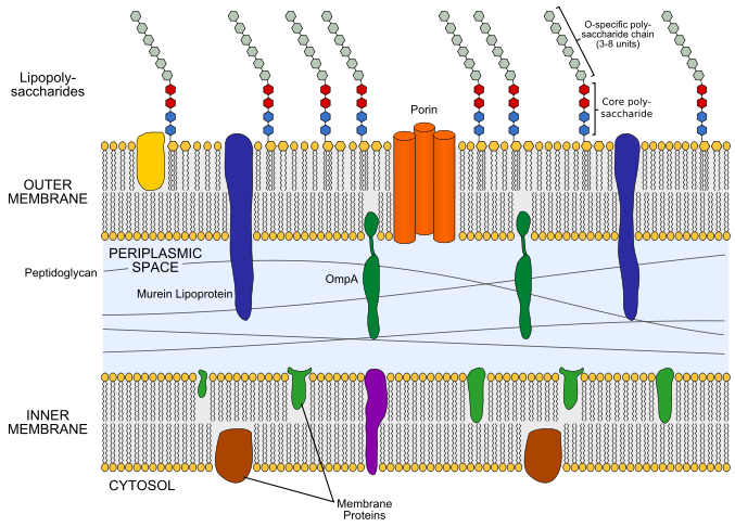
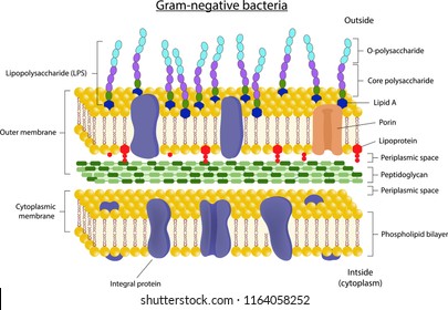
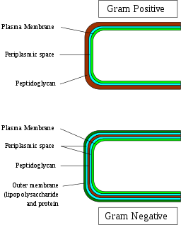
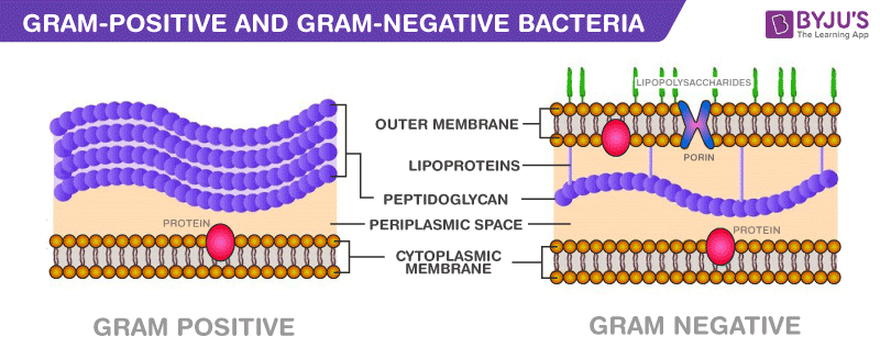
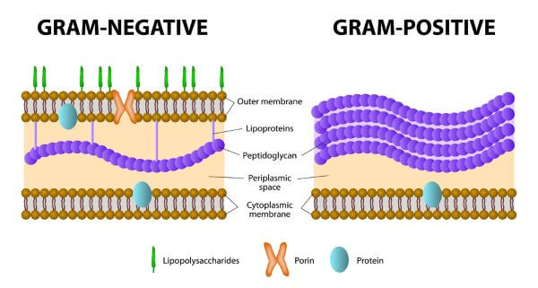
June 2, 2020 - Most bacteria are classified into two broad categories: Gram positive and Gram negative. These categories are based on their cell wall composition and reaction to the Gram stain test. The Gram staining method, developed by Hans Christian Gram, identifies bacteria based upon the reaction of ... August 19, 2021 - The cell membrane of Gram-positive ... of Gram-negative bacteria. Some examples of Gram-positive bacteria include Streptococcus, Staphylococcus, and Clostridium botulinum (botulism toxin). Gram-positive bacteria have a greater volume of peptidoglycan (a polymer of amino acids and sugars that create the cell wall of all bacteria ... March 1, 2021 - The Gram-negative cell wall is composed of an outer membrane, a peptidoglygan layer, and a periplasm. January 3, 2021 - Because of the nature of their cell wall, Gram-negative bacteria stain pink after Gram staining. The Gram-negative cell wall consists of 2-3 interconnected layers of peptidoglycan surrounded by an …
a) Diagram of a gram-negative cell wall. (b) Electron ...
Like they pick up a bit of dna from somewhere else and start making the wrong one, would the cell just fall apart?

Hi....✌➡ 2 Differences between gram positive and gram ...
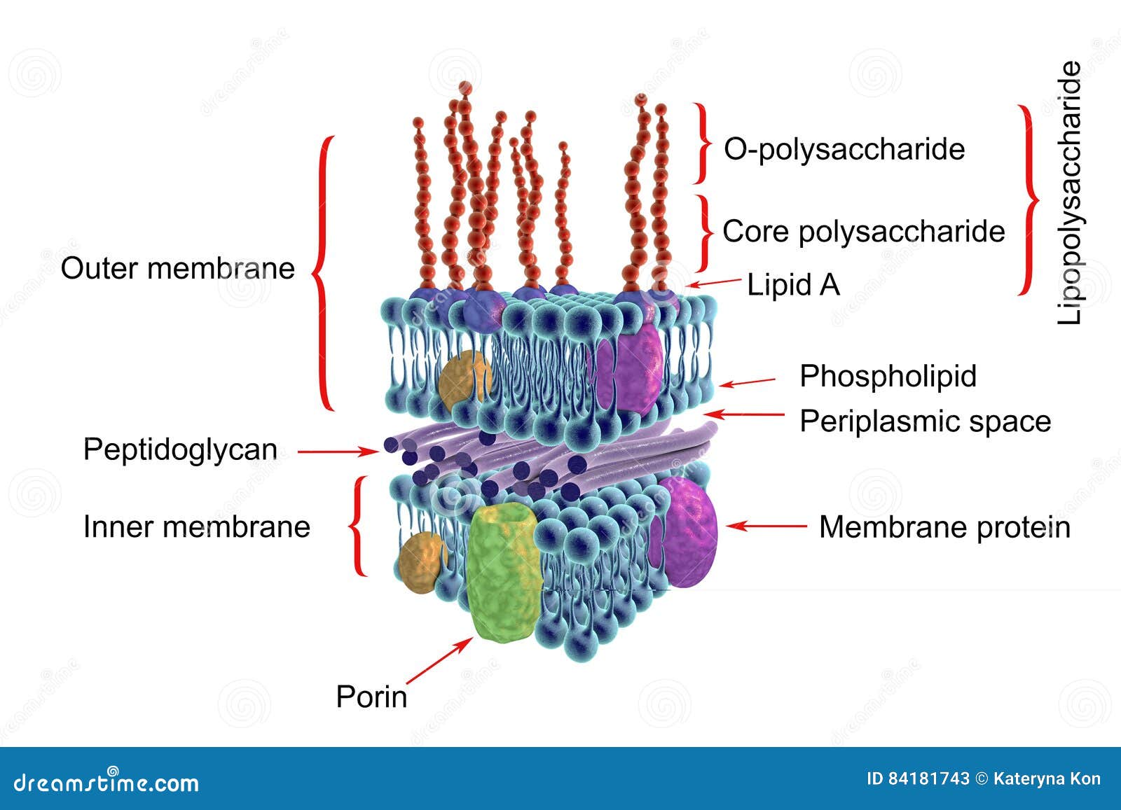
February 15, 2021 - Figure 1. Upper image. Gram-negative bacterial cell wall. Lower image. Lipopolysaccharide Cytoplasmic membrane: Surrounds the cytoplasm of Gram-positive and Gram-negative bacteria and regulates the passage of molecules in and out of the bacteria. It is similar to the one found in the mammalian ...
VetBact
August 15, 1999 - The core oligosaccharide and lipid A moieties are not shown but would be at the bottom of the diagram. ... Negative-stained n-MVs which have been isolated and purified from P. aeruginosa as previously described (32). Bar = 250 nm. ... Thin section of an unidentified gram-negative bacterium ...

Cell wall schematic of (A) Gram-positive and (B) Gram ...
It is important to note that not all bacteria have a cell wall. Having said that though, it is also important to note that most bacteria (about 90%) have a cell wall and they typically have one of two types: a gram positive cell wall or a gram negative cell wall.
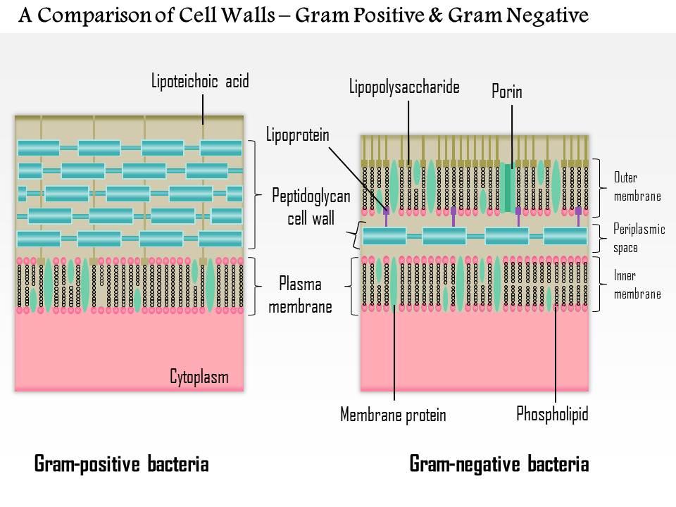
0614 A Comparison Of The Cell Walls Gram Positive And Gram ...
Hi all basically after stupidly using an unclean vibrator last june i’ve had recurring vaginal pain after penetration that metronidazole only seems to mask. I had awful pain, throbbing, vaginal aching, and rotten smell. metronidazole seemed to calm everything down a bit. swabs were clear however. during this one of my bartholin glands became inflamed for WEEKS- didn’t turn into a cyst but i had pain on arousal which eventually cleared out on its own. basic swabs between june - august from the...
Bacteria: Cell Walls – General Microbiology
what would happen after the basic stain, alcohol de-stain, and counterstain? the basic stain would still be attracted to the negatively charged cell membrane correct? but then the stain would not be retained due to lack of cell wall (peptidoglycan) after the de-stain? so then the end result would be that the cell would be stained the color of the counterstain? thanks in advance!
What is the Difference Between Gram Positive and Gram ...
Download scientific diagram | Schematic structure of Gram-positive and Gram-negative cell walls. Gram-positive cell walls contain only one lipid plasma membrane and a thick peptidoglycan layer interlinked with teichoic and lipoteichoic acids, whereas Gram-negative bacteria have an inner and ...

Bacteria basics – Part 2 – ReAct
Most procaryotes have a rigid cell wall. The cell wall is an essential structure that protects the cell protoplast from mechanical damage and from osmotic rupture or lysis. Procaryotes usually live in relatively dilute environments such that the accumulation of solutes inside the procaryotic ...
Lipid A and Bacterial Lipopolysaccharides, Gram-negative ...
Gram-negative cell walls are strong enough to withstand ∼3 atm of turgor pressure (40), tough enough to endure extreme temperatures and pHs (e.g., Thiobacillus ferrooxidans grows at a pH of ≈1.5) and elastic enough to be capable of expanding several times their normal surface area (41).

DIFFERENCE BETWEEN GRAM NEGATIVE AND GRAM POSITIVE CELL WALL ...
Gram-negative bacteria are surrounded by a thin peptidoglycan cell wall, which itself is surrounded by an outer membrane containing lipopolysaccharide. Gram-positive bacteria lack an outer membrane but are surrounded by layers of peptidoglycan many times thicker than is found in the Gram-negatives.

Gram-Positive vs. Gram-Negative Bacteria • Microbe Online
August 10, 2021 - Gram-positive cell wall. The Gram-positive cell wall is thick (15–80 nm) and more homogenous than that of the thin (2 nm) Gram-negative cell wall. The Gram-positive cell wall contains large amount of peptidoglycan present in several layers that constitutes about 40–80% of dry weight of ...

Gram-negative cell Diagram | Quizlet

Bacterial Pathogens Research
Cell Wall - Bacteria
Cell wall Images, Stock Photos & Vectors | Shutterstock
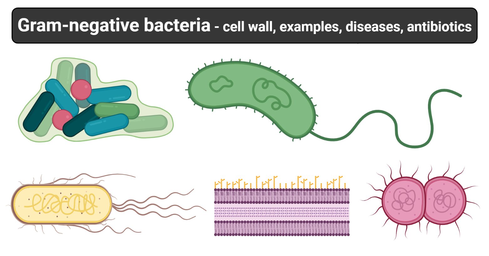
Gram-negative bacteria- cell wall, examples, diseases ...
Difference between Gram-Positive and Negative Bacteria - Gram ...

Gram negative cell wall Diagram | Quizlet
Gram Positive and Gram Negative Bacteria - Structures ...

Difference between Gram Positive and Gram Negative Bacteria ...
Major Difference Between Gram-Positive and Gram-Negative Bacteria
Gram-Positive vs. Gram-Negative | Biology Dictionary

25 Differences between gram positive and gram negative bacteria

Gram-Negative Bacterial Cell Wall, Unit 1 Diagram | Quizlet
Bacterial Endotoxin

Pathogen Recognition and Innate Immunity: Cell

Bacterial Cell Structure Wall Gram-positive Bacteria Gram ...
Gram positive cell Images, Stock Photos & Vectors | Shutterstock

Bacterial Cell Wall. The differences between Gram-positive ...
Schematic diagram comparing the cell envelopes of (a) gram ...

Bacterial Cell Wall: Types, Composition and Function ...
Gram Negative Bacteria Cell Wall Stock Illustrations – 13 ...

Peptidoglycan Structure, Biosynthesis and Function

Cell Wall Structure of Gram-negative Bacteria for Example ...
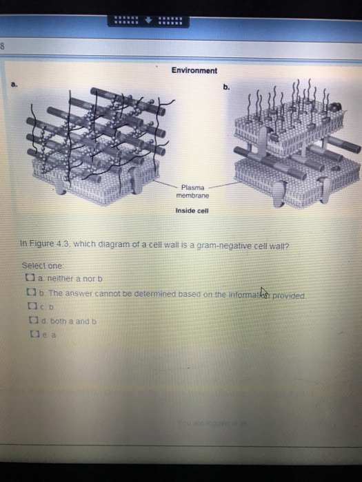
Solved In figure 4.3 which diagram of a cell is a gram ...
Through the wall: extracellular vesicles in Gram-positive ...

2.png

Solved er 4 Multiple-Choice Question 27 Part A Environment ...
0 Response to "38 gram negative cell wall diagram"
Post a Comment