39 compound microscope ray diagram
Hi all, I saw a [post earlier today](https://www.reddit.com/r/ufo/comments/e6td91/chris_cogswell_discusses_the_metamaterials_and/) about Chris Cogswell discussion regarding TTSA's "metamaterials" (massive emphasis on the quotation marks) and how to prove whether they're made by humans or not. Reading some of his comments, I realize he is missing information that I have access to and it's not fair to him or anyone else in the community. I was reached out to by an American scientist who owns a ve... Compound Microscope - Diagram (Parts labelled), Principle and Uses. Also called as binocular microscope or compound light microscope, it is a remarkable magnification tool that employs a combination of lenses to magnify the image of a sample that is not visible to the naked eye.
Diagram representation of scanning probe microscope. X-ray microscope - uses X-rays in place of visible light and is used to look inside small objects and structures. Compound microscope diagram. Facebook Twitter Google+ Pinterest Linkedin.

Compound microscope ray diagram
An animated presentation showing you how to draw ray diagrams (using simple lens rules) for a compound microscope. This shows how to determine the position and magnification of the final (virtual) image. Thursday, 10 November 2011. Ray Diagram for Compound microscope. Ray Diagram of Normal Eye, Short-sightedness, Long-sightedness and their correction. Diagram of a compound microscope. A compound microscope uses a lens close to the object being viewed to collect light (called the objective lens) which focuses a real Additionally, methods such as electron or X-ray microscopy use a vacuum or partial vacuum, which limits their use for live...
Compound microscope ray diagram. Sai Vara Prasad answered this. this is the diagram of compound micro scope. REGARDS A compound microscope is a type of microscope that uses two sets of lenses to magnify the image under the microscope. It has an objective lens that has a resolution of 4x,10x, 40x, 100x, and an eyepiece of resolution of 10x. Ray diagram of phase contrast microscope. When we use a usual biology class compound microscope, we need to focus the stage (place the object) to the "correct" position. It is just in that sweet spot that the image is sharp and clear. So we are deciding the object distance. Here is the ray diagram of a compound microscope. A compound microscope is a laboratory instrument used to magnify the image of a small object; usually objects that cannot be seen by the naked eye. Principles of compound microscope. When a minute object is placed beyond the focus of the objective lens, a highly magnified object is formed at a...
Compound Microscope and its Magnification. It is an optical device used to observe very small objects like bacteria, cells, gas molecules and section When ever high magnification is desired, a compound microscope is used. It consists of two convex lenses, one placed before the object called Objective... Working Principle of Compound Microscope. Parts of compound microscope. Applications and Uses. I'm having some difficulty grasping question 8(b)(ii) of [this IB past paper](http://www.uplifteducation.org/cms/lib01/TX01001293/Centricity/Domain/273/TEST%20OPTIONS%202013.pdf): In a compound microscope, the distance between the lenses is 18.1 cm. The focal length of the eyepiece lens is 3.8 cm. The intermediate image forms 14.8 cm from the objective lens, i.e. 3.3 cm from the eyepiece lens. From part (i), we know the final image forms 25.1 cm from the eyepiece lens. Part (ii) states that th... (ii) Why must both the objective and the eye-piece of a compound microscope have short focal lengths? Draw a neat labeled ray diagram of a compound microscope.
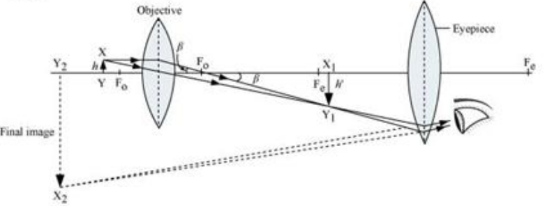
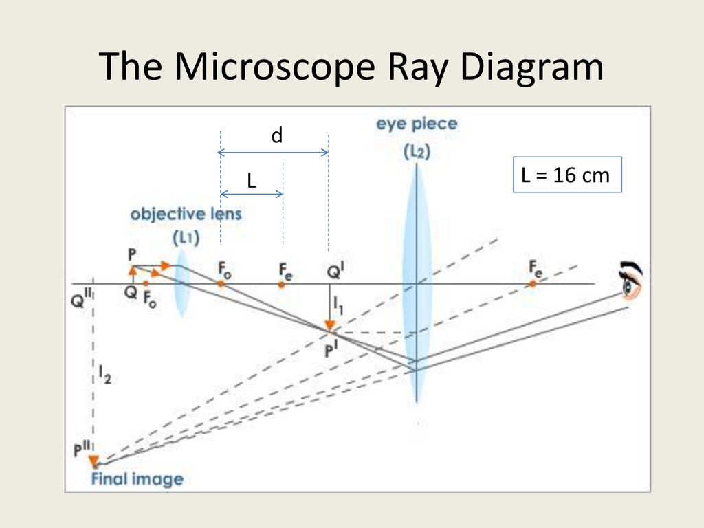
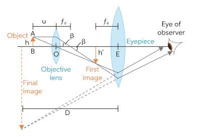
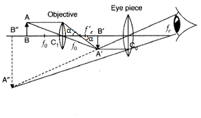
A schematic diagram of a compound microscope is shown in Fig. The lens nearest the object, called the objective, forms a real, inverted, magnified image of the object. This serves as the object for the second lens, the eyepiece, which functions essentially like a simple microscope or magnifier... The compound microscope and ray diagrams. It is a rectangular metal plate fitted to the vertical rod. Draw a labelled ray diagram to show the formation of the image by a compound microscope. A simple microscope uses the optical power of single lens or group of lenses for magnification. These microscopes provide better magnification than light microscopes. Compound Microscope Diagram. The compound microscope uses light for illumination. Some compound microscopes make use of natural light, whereas others have an illuminator attached to the base. Compound Microscope: Uses a lens close to the object that's being viewed to collect light (this is called an objective lens) This creates a real image of the object inside the microscope (image 1 of ray diagram), Then, that image is magnified by the second lens (or group of lenses) called the eye piece.
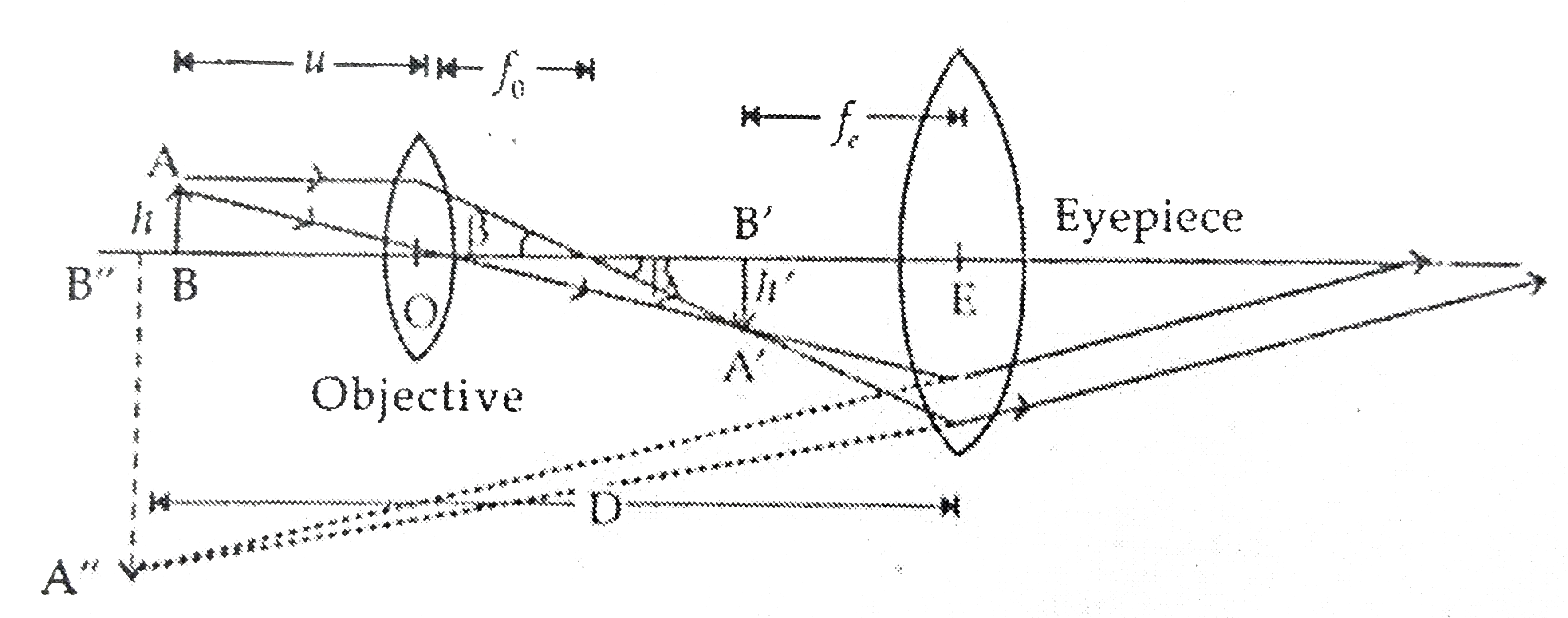
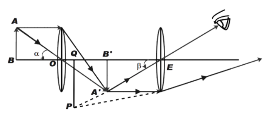
Here are two ray diagrams for compound microscope, the first one proposed by the book, and the second one recommended by the teacher Both diagrams are 'correct' (meaning they both correctly locate the virtual image), but the second diagram is more in the spirit of ray tracing.

(a)Draw a ray dia. (a) A microscope is a device used to see magnified image of very small things which, a compound microscope consists of two convex lenses namely eyepiece and objective, objective lens makes enlarged the real image of object which acts as an object for eyepiece which...

Modern compound microscopes are designed to provide a magnified two-dimensional image that Figure 1 - The Microscope Optical Train. Most microscopes provide a translation mechanism Simple ray diagrams are sufficient to explain many important aspects of microscopy including...

Extremely helpful video for students who are appearing for board exams.How to draw perfect Concave & Convex lenses with pencil and Compasshttps...
Explore microscope parts and functions. The compound microscope is more complicated than just a microscope with more than one lens. With Labeled Diagram and Functions. How does a Compound Microscope Work?
Videos for Microscope and Telescope The astronomical telescope ray diagram What is Compound Microscope A compound microscope has two lenses. One is called eyepiece and the other is called...

(a) Image formation by a compound microscope : A schematic diagram of a compound microscope is shown in Fig. <br> <img src="https (i) Draw a neat labelled ray diagram of a compound microscope. Explain briefly its working. <br> (ii) Why must both the objective and the eye-piece of a...
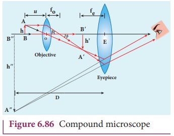
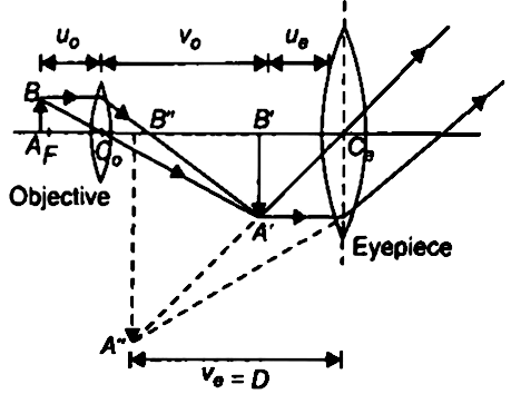
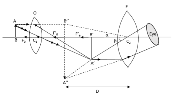
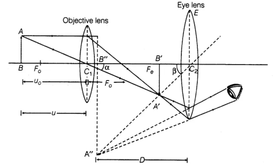
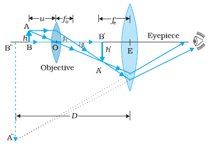
In this case, the objective lens O of the compound microscope forms a real, inverted and enlarged image A'B' of the object. Now A'B' acts as an object for the eyepiece E, whose position is adjusted so that A'B' lies between optical centre C2 and the focus fe' of eyepiece.

Diagram of a Compound Microscope. Article Shared by. ADVERTISEMENTS A bright-field or compound microscope is primarily used to enlarge or magnify the image of the object that is (ii) The angular aperture, i.e., the angle between the most divergent rays passing through the lens and optical...
Ray Diagram - Compound Microscope. How to draw a compound microscope diagram..Подробнее. Compound microscopeПодробнее. Compound microscopeПодробнее. Compound microscopeПодробнее.

Learn to draw Compound Microscope Diagram (Final Image at least distance of distinct vision "D") within 3 mins. Background ... This video tries to instruct method to draw ray diagram of an Compound Microscope forming image at near point, It deals with ...
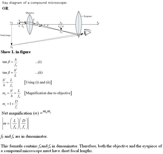
A compound microscope consists of objective lens and eyepiece mounted coaxially (having a common axis) the position of the eyepiece is so adjusted that the image lies within the focus of the eye lens. Working The ray diagram given below gives the principle of a compound microscope.
a compound microscope is the combination of two convex lenses the objective and the eyepiece. this lens with a small focal lenth this lens is placed close to the object. Earn +20 pts. Q: Describe compound microscope with ray diagram?
Ray diagram of a compound microscope.When the final image is formed at the least distance of distinct vision,For the image formed at infinity, ue = feand By making focal length of the objective small, the magnifying power can be increased.
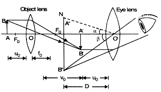
Diagram of a compound microscope. A compound microscope uses a lens close to the object being viewed to collect light (called the objective lens) which focuses a real Additionally, methods such as electron or X-ray microscopy use a vacuum or partial vacuum, which limits their use for live...

Thursday, 10 November 2011. Ray Diagram for Compound microscope. Ray Diagram of Normal Eye, Short-sightedness, Long-sightedness and their correction.

An animated presentation showing you how to draw ray diagrams (using simple lens rules) for a compound microscope. This shows how to determine the position and magnification of the final (virtual) image.


0 Response to "39 compound microscope ray diagram"
Post a Comment