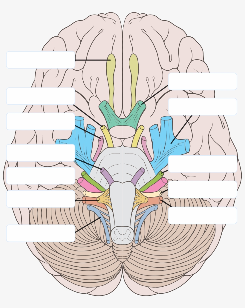
39 blank cranial nerve diagram
Fill-in-the-Blank Cranial Nerve Nuclei Diagram. Resident PRITE Review. LLU Residents using Brain Stem Diagram during PRITE Neurology Review. Resident PRITE Review. LLU Residents using Brain Stem Diagram during PRITE Neurology Review. View fullsize. Diseases of the Neuromuscular Junction Concept Map - posted on instagram @minipsychmd, post here. Dec 06, 2021 · The cranial nerves are named after the body parts that they serve and are also assigned Roman numerals based off their location from front to back. 3D Animation Best viewed with Netscape Navigator BrainstemCranial Nerves Anterior View Blank Diagram Anterior View Complete Diagram Lateral View Blank Diagram Lateral View Complete Diagram Spinal Cord.
Circular muscles of the iris are controlled by the (blank) nervous system and cause the pupil to (blank). Radial muscles of the iris are controlled by the (blank) nervous system and cause the pupil to (blank). The circular muscles are controlled specifically by the (blank) cranial nerve.
Blank cranial nerve diagram
The following links should either display a PDF fill-in the blank document in the browser or present an option to download the file to your computer. In either case, you should be able to print any of these files. It is recommended that they be printed on an inkjet printer for maximum quality. The student will gain the benefit of learning names ... Labeled brain diagram. First up, have a look at the labeled brain structures on the image below. Try to memorize the name and location of each structure, then proceed to test yourself with the blank brain diagram provided below. Labeled diagram showing the main parts of the brain. Complete Diagrams. Brain Ventricles: 3D Animation (Best viewed with Netscape Navigator) Brainstem/Cranial Nerves. Anterior View (Blank Diagram) Anterior View (Complete Diagram) Lateral View (Blank Diagram) Lateral View (Complete Diagram) Spinal Cord: Cross Section. Blank Diagram.
Blank cranial nerve diagram. Sep 13, 2021 · This human anatomy module is about the cranial nerves. It consists of 15 vector anatomical drawings with 280 anatomical structures labeled. It is intended for the use of medical students working on human anatomy, student nurses, physiotherapists, electro-radiological technicians and residents – especially those working in neurology, neurosurgery, otolaryngology – and for any physician ... The functions of the cranial nerves are sensory, motor, or both: Sensory cranial nerves help a person to see, smell, and hear. Motor cranial nerves help control muscle movements in the head and neck. 1.1 Attachments of cranial nerves, anterior view page 8 1.2 Attachments of cranial nerves, lateral view 9 1.3 Ganglia and nuclei 12 2.1 Cranial nerve motor nuclei 23 3.1 Corticonuclear pathways 26 4.1 Trigeminal sensory system 34 7.1 Trigeminal nerve 51 8.1 Ophthalmic nerve 53 9.1 Maxillary nerve 57 10.1 Mandibular nerve 61 11.1 Facial nerve ... External anatomy of the eye diagram unlabeled.If you would like a large unwatermarked image for your web page or. 12 Best Images of Anatomy Human Ear Diagram Worksheet - Blank Ear Diagram Human Eye Diagram Unlabeled and General and Special Senses Worksheet See 12 Best Images of Anatomy Human Ear Diagram Worksheet.
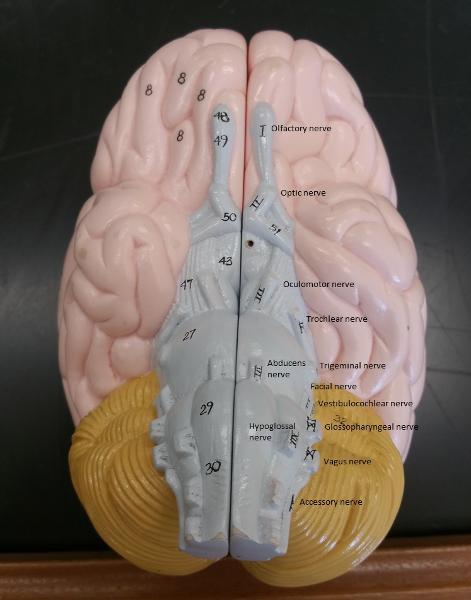
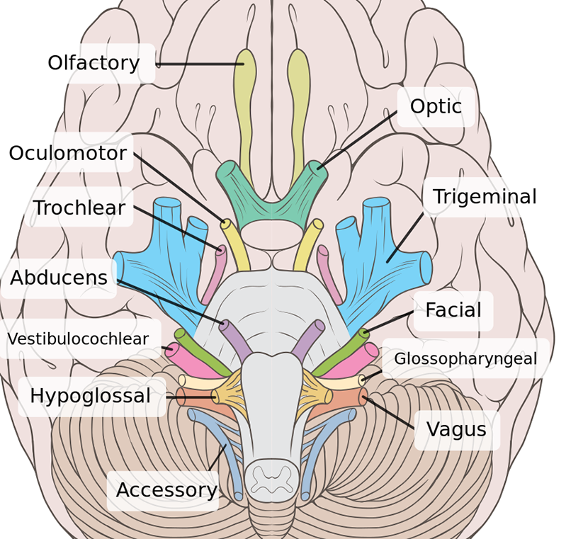
Each of the 12 cranial nerves is represented students color and number each nerve in both brains. All the functions are carried out without a single glitch and before you even bat an eyelid. Learn all the parts of the human heart by memorizing this free printable human heart diagram. 16 Unlabeled Brain Diagram Pictures. Cranial Nerve 5. Motor and sensory nerve-Trigeminal Nerve-Carries sensory information from most of the head, neck, sinuses, and face. The cranial nerves are in contrast to spinal nerves, which emerge from segments of the spinal cord. Contents. 1 Anatomy. 1.1 ... The 12 cranial nerves are pairs of nerves that start in different parts of your brain. Learn to explore each nerve in a 3-D diagram.
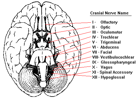
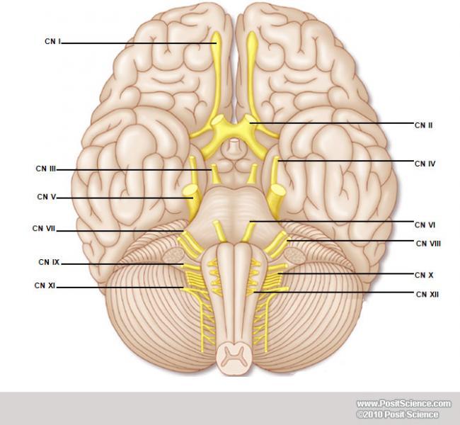
Cranial Nerve Major Functions Assessment Cranial Nerve I Olfactory Sensory Smell Smell—coffee, cloves, peppermint Cranial Nerve II Optic Sensory Vision Visual acuity—Snellen chart (cover eye not being examined) Test for visual fields Examine with ophthalmoscope Cranial Nerve III Oculomotor Sensory and ... with more related ideas as follows unlabeled pelvis bone anatomy, vertebrae diagram unlabeled and cranial nerves labeling quiz. We hope these Blank Anatomy Worksheets photos collection can be a guide for you, give you more references and also help you get what you search. A detailed overview of the 12 pairs of cranial nerves, including one diagram. 1 page document. Great for revision of cranial nerves in courses such as speech pathology or communication sciences. ... 2 page worksheet includes cranial nerve fill-in-the-blank table with mix and match rows that are out of order (not in numerical order I through XII ... Cranial nerves blank diagram. The following diagram is provided as an overview of and topical guide to the human nervous system. Their numerical order 1-12 is determined by their skull exit location rostral to caudal. While we talk about Human Anatomy Labeling Worksheets we already collected several similar pictures to inform you more.
Neuroscience for Kids - Cranial Nerves
Blank Cranial Nerve Diagram via. In our website, we are people that are very admire original idea from every one, without exception! we make sure to keep the original photos without changing anything including the watermark. Every pictures gallery we publish are always carrying the original website link where we found it below each images.

Cranial Nerves – Biology for Everybody
A topographical anatomy of the brain showing the different levels (encephalon, diencephalon, mesencephalon, metencephalon, pons and cerebellum, rhombencephalon and prosencephalon) as well as a diagram of the various cerebral lobes (frontal lobe, occipital, parietal, temporal, limbic and insular).
CN Flashcards | Chegg.com
Nov 15, 2016 - Shows pictures of a sheep and a human brain. Each of the 12 cranial nerves is represented, students color and number each nerve in both ...
The Cranial Nerves: 9780982748510: Medicine & Health Science ...
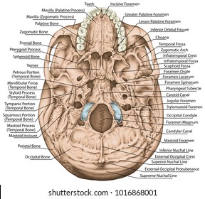
Learn the major cranial bone names and anatomy of the skull using this mnemonic and labeled diagram. Sutures connect cranial bones and facial bones of the skull. Develop a good way to remember the cranial bone markings, types, definition, and names including the frontal bone, occipital bone, parieta

What's Behind that Smile: Using Analogies, Facial Expressions ...
Blank Diagram Complete Diagram. Brain Ventricles: Anterior View Blank Diagram Complete Diagram. Brain Ventricles: Lateral and Superior Views Blank Diagrams Complete Diagrams Brain Ventricles: 3D Animation The Homunculus . Brainstem/Cranial Nerves Anterior View (Blank Diagram) Anterior View (Complete Diagram) Lateral View (Blank Diagram)

Cranial Nerve
30 Sept 2017 ... Cranial Nerves (Anatomy and Functions); explained beautifully in an illustrated and interactive way. Click and start learning now!
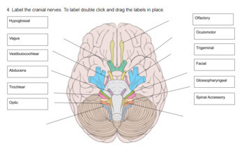
Activity 3: Cranial Nerves and Identifying and Testing ...
These nerves are paired and present on both sides of the body. They are mainly responsible for facilitating smell, vision, hearing, and movement of muscles. Cranial nerves are concerned with the head, neck, and other facial regions of the body. Cranial nerves arise directly from the brain in contrast to spinal nerves and exit through its foramina.
Cranial nerve Images, Stock Photos & Vectors | Shutterstock
The cranial nerves are 12 pairs of nerves that can be seen on the ventral (bottom) surface of the brain. Some of these nerves bring information from the sense organs to the brain; other cranial nerves control muscles; other cranial nerves are connected to glands or internal organs such as the heart and lungs.
Cranial nerves quizzes and labeling exercises | Kenhub
Cranial Nerves Quiz for Anatomy & Physiology Class. This cranial nerves exam will test your knowledge on all the cranial nerves that you will have to know for an exam in Anatomy & Physiology. This cranial nerves quiz will ask you about the function and name of each nerve. 1. There are 14 pairs cranial nerves. *. True. False.
ventricles of the brain not labeled - Clip Art Library
The facial nerve is also known as the seventh cranial nerve (CN7). This nerve performs two major functions. It conveys some sensory information from the tongue and the interior of the mouth.
Neuroanatomy and physiological workbook
Oct 28, 2021 · If it’s helpful for you, you can also download the labeled cranial nerves diagram and use it to make notes. Download PDF Worksheet (blank) Download PDF Worksheet (labeled) Now let’s look at some different type of cranial nerve quizzes you can take. Interactive quizzes

Download Free Vectors, Cliparts, Images of Cranial nerve ...
Jun 24, 2021 · The sensory cranial nerves are involved with the senses, search as sight, smell, hearing, and touch. Whereas the motor nerves are responsible for controlling the movements and functions of muscles and glands, cranial nerves supply sensory and motor information to areas of the head and neck. One nerve, the vagus nerve, extends beyond the neck to ...

JaypeeDigital | eBook Reader
Start studying Cranial Nerve Chart Fill in the Blank. Learn vocabulary, terms, and more with flashcards, games, and other study tools.
/GettyImages-141483691-4cc225237a5945f8ab949d936f52c48e.jpg)
Cranial Nerves: Anatomy, Function, and Treatment
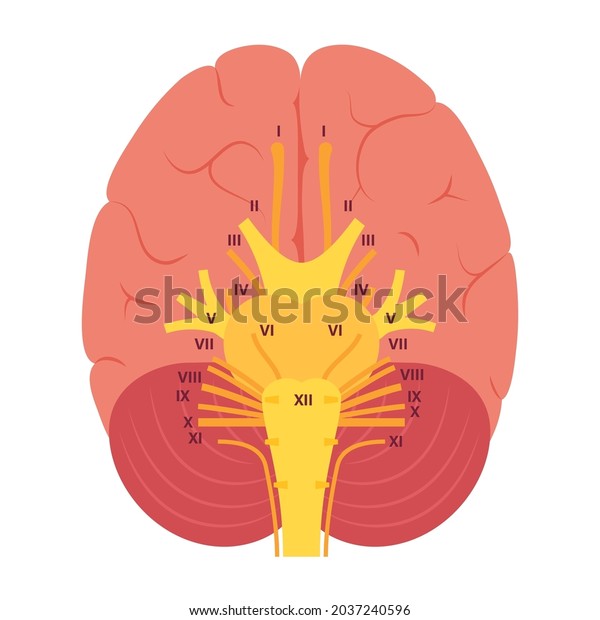
The twelve cranial nerves, in order from I to XII are: olfactory nerve, optic nerve, oculomotor nerve, trochlear nerve, trigeminal nerve, abducens nerve, facial ...

Nervous about nerves? - a review of cranial nerves ...
Complete Diagrams. Brain Ventricles: 3D Animation (Best viewed with Netscape Navigator) Brainstem/Cranial Nerves. Anterior View (Blank Diagram) Anterior View (Complete Diagram) Lateral View (Blank Diagram) Lateral View (Complete Diagram) Spinal Cord: Cross Section. Blank Diagram.

Human Brain / Cranial Nerves Stock Illustration ...
Labeled brain diagram. First up, have a look at the labeled brain structures on the image below. Try to memorize the name and location of each structure, then proceed to test yourself with the blank brain diagram provided below. Labeled diagram showing the main parts of the brain.
Cranial Nerves Virtual Lab
The following links should either display a PDF fill-in the blank document in the browser or present an option to download the file to your computer. In either case, you should be able to print any of these files. It is recommended that they be printed on an inkjet printer for maximum quality. The student will gain the benefit of learning names ...

Cranial Nerves Diagram | Quizlet
How to Learn the 12 Cranial Nerves

nervoussystemdiagrams
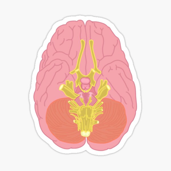
Cranial Nerves Stickers | Redbubble

Hypoglossal Nerve Greeting Cards | Fine Art America
Cranial nerves quizzes and labeling exercises | Kenhub

Nervous about nerves? - a review of cranial nerves - maidoodles

Illustrations and diagrams of the 12 pairs of cranial nerves ...
Cranial Nerves Diagram Brain Structure Medical Stock Vector ...
What are the 12 cranial nerves? Functions and diagram

Cranial Nerves Identification Quiz

HyperBrain Syllabus Chapter 1

Cranial Nerves Anatomy

Cranial nerve
Vestibulocochlear nerve (CN VIII): Anatomy and pathway | Kenhub

JaypeeDigital | eBook Reader
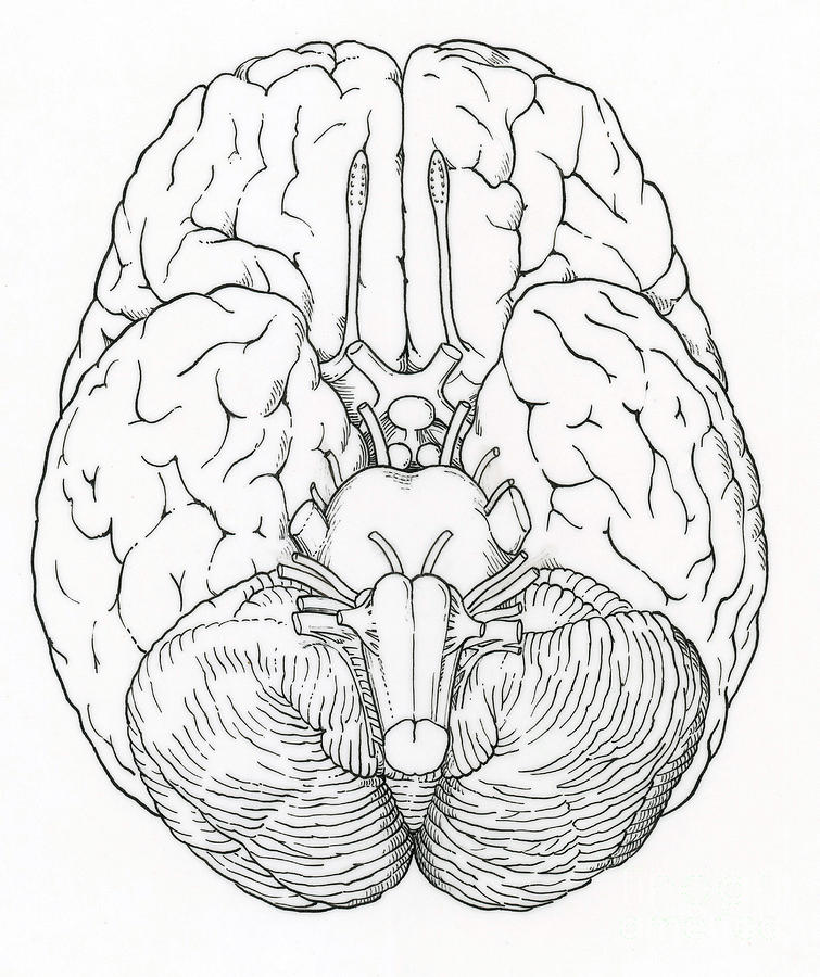
Illustration Of Cranial Nerves Photograph by Science Source
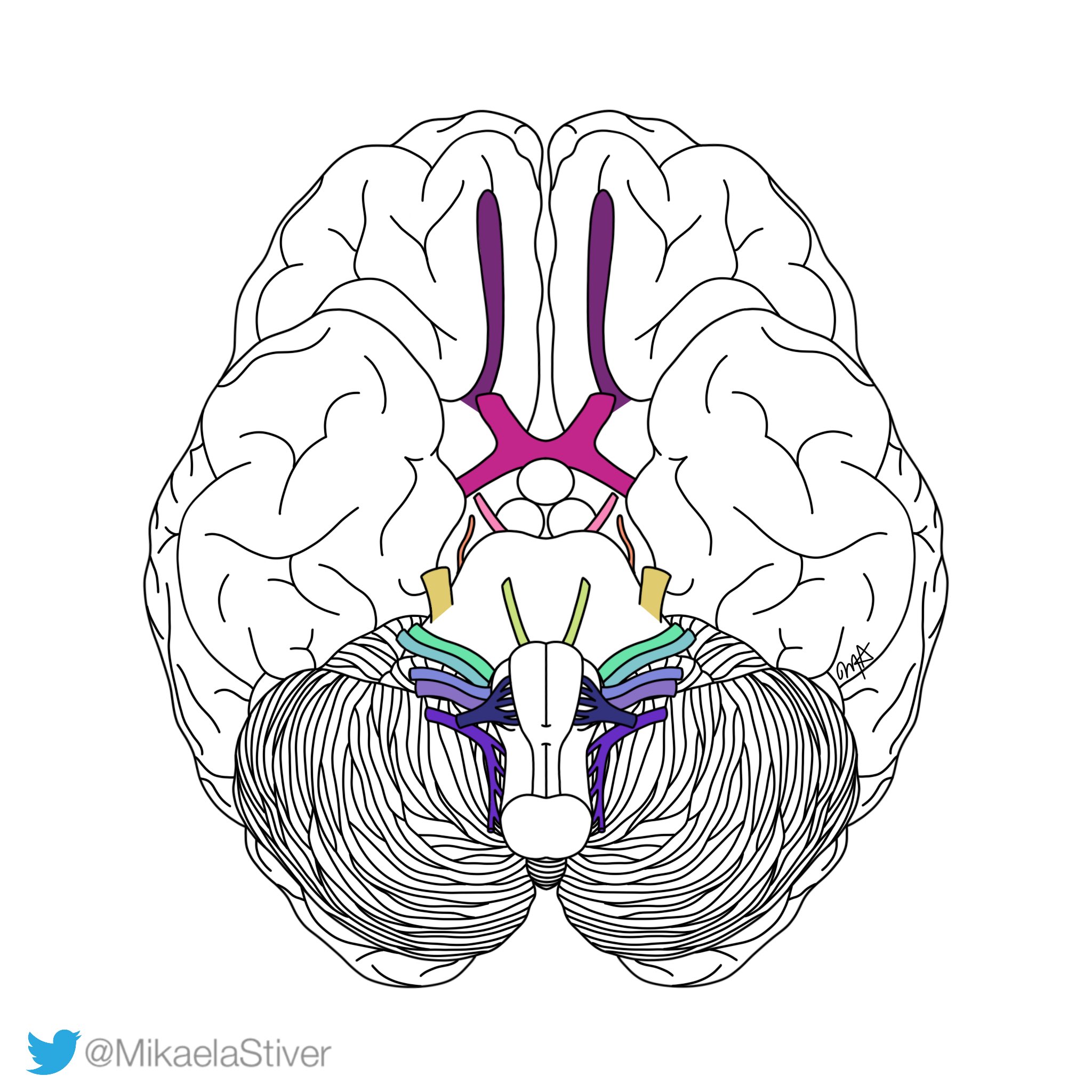
Mikaela Stiver on Twitter: "Behold!! The cranial nerve ...
/GettyImages-141483691-4cc225237a5945f8ab949d936f52c48e.jpg)
Cranial Nerves: Anatomy, Function, and Treatment

Cranial Nerves - Anatomy and Functions
Nerves Clipart Nervous Boy - 12 Cranial Nerves Blank PNG ...
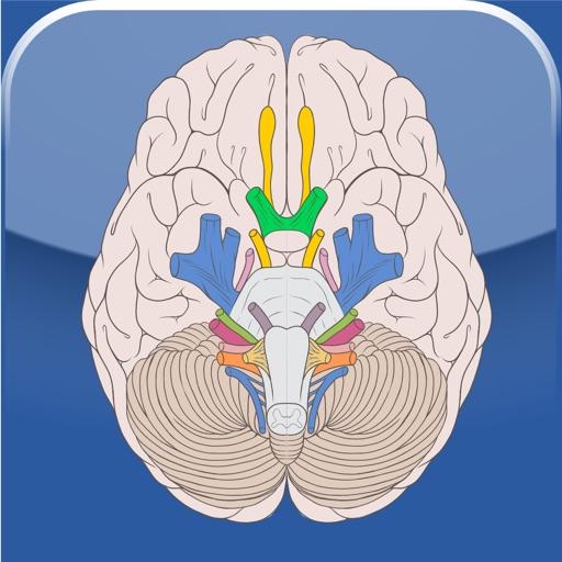
Cranial Nerves - Apps on Google Play
0 Response to "39 blank cranial nerve diagram"
Post a Comment