39 diagram of the back muscles
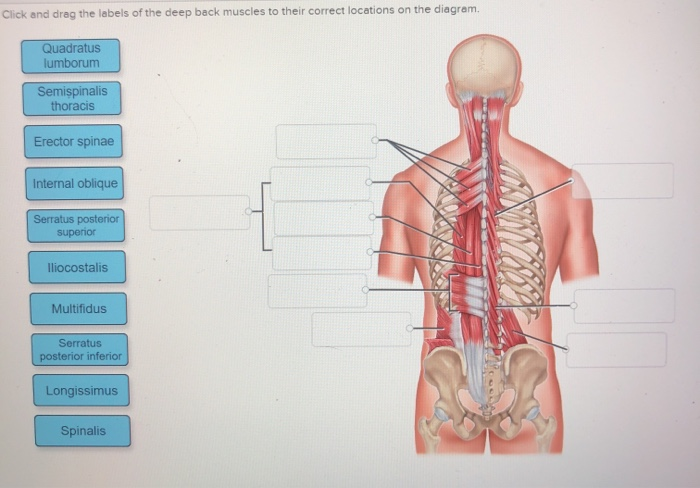
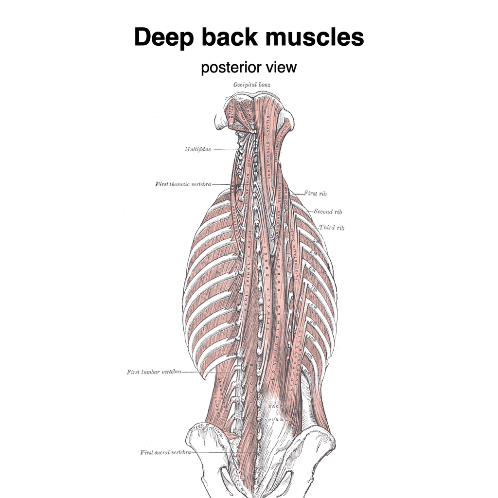
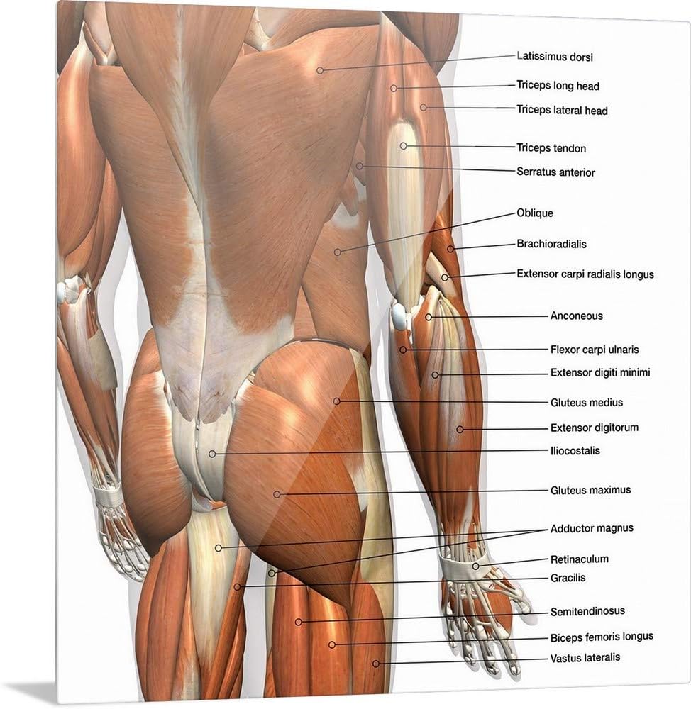
The muscles of the back can be arranged into 3 categories based on their location: superficial back muscles, intermediate back muscles and intrinsic back muscles.The intrinsic muscles are named as such because their embryological development begins in the back, oppose to the superficial and intermediate back muscles which develop elsewhere and are therefore classed as extrinsic muscles. Three types of back muscles that help the spine function are extensors, flexors and obliques. The extensor muscles are attached to back of the spine and enable standing and lifting objects. These muscles include the large paired muscles in the lower back, called erector spinae, which help hold up the spine, and gluteal muscles.
We hope this picture Anatomy Of Back Muscles Diagram can help you study and research. for more anatomy content please follow us and visit our website: www.anatomynote.com. Anatomynote.com found Anatomy Of Back Muscles Diagram from plenty of anatomical pictures on the internet. We think this is the most useful anatomy picture that you need.

Diagram of the back muscles
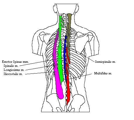
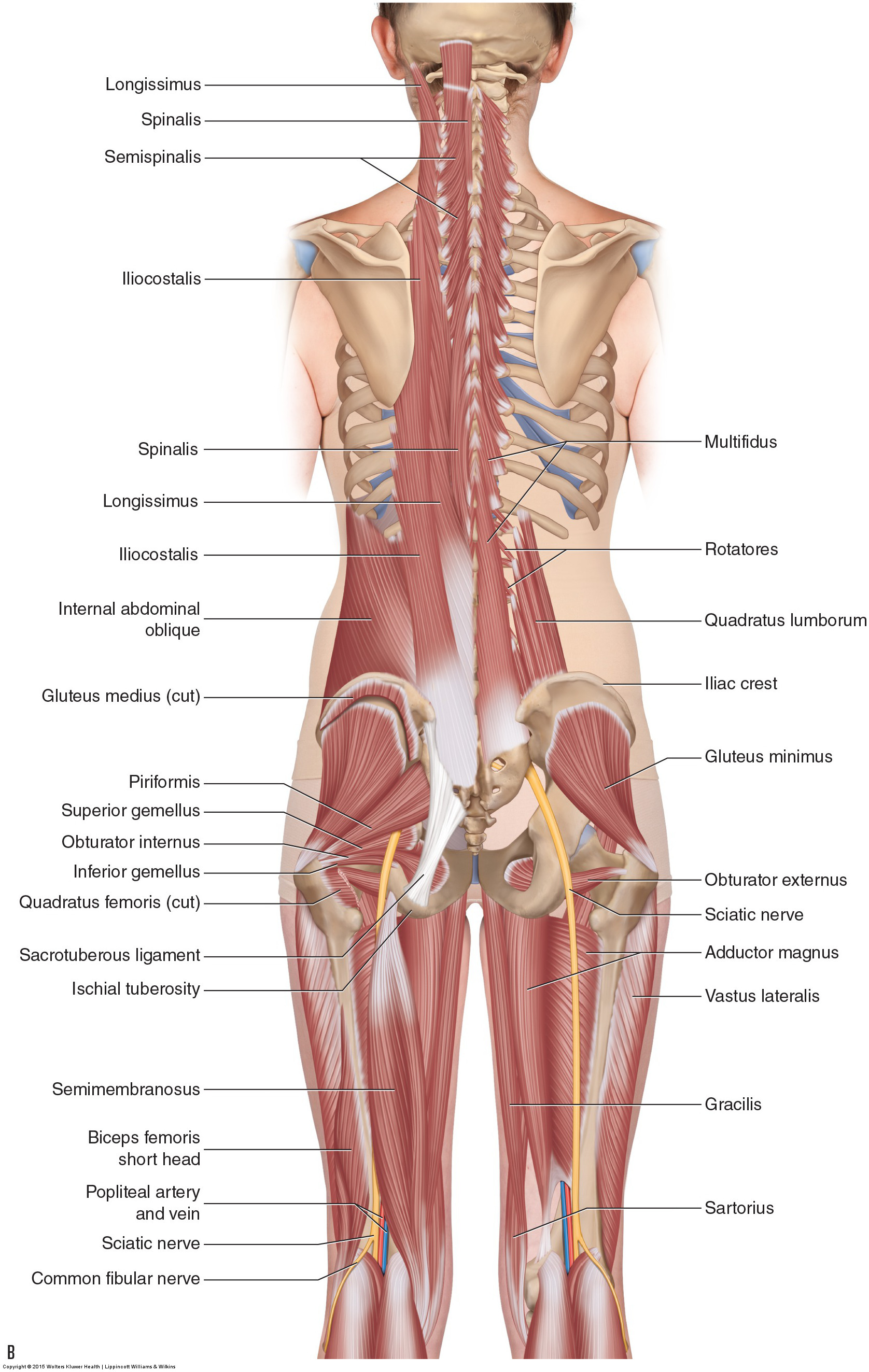
740 lumbar spine anatomy diagram stock photos, vectors, and illustrations are available royalty-free. See lumbar spine anatomy diagram stock video clips. of 8. spinal vertebrae bone spine vertebra toracica spinal cord spine structure back diagram spine sections spinal cord vertebrae spinal structure health diagram. Try these curated collections. The muscles of your back support your spine, attach your pelvis and shoulders to your trunk, and provide mobility and stability to your trunk and spine. The anatomy of your back muscles can be complex. There are several different layers of muscles in your back that are often pulling in different and various directions. Muscles found in the deep group include the spinotransversales, erector spinae (composed of the iliocostalis, longissimus, and spinalis), the transversospinales, and the segmental muscles. "The best way to strengthen back muscles is in a static position. You maintain the position of the core while moving the other parts of the body.".
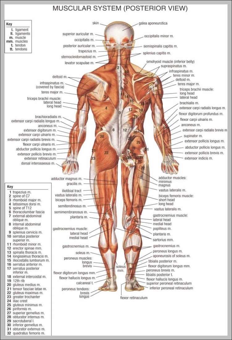
Diagram of the back muscles. Back anatomy. The back is the body region between the neck and the gluteal regions. It comprises the vertebral column (spine) and two compartments of back muscles; extrinsic and intrinsic. The back functions are many, such as to house and protect the spinal cord, hold the body and head upright, and adjust the movements of the upper and lower limbs. Back Muscles, Back Muscle Diagram. Creatine is now proving to be one of the most potent muscle growth accelerators giving excellent muscle mass increase and phenomenal strength increases order yours today. Creatine Research More Than a Sports Supplement Read More…. Some of the links in the post above are "affiliate links.". See Back Muscles and Low Back Pain. Nerves in your lower back. Five pairs of lumbar spinal nerves labeled L1 to L5 branch off your spinal cord and exit through small holes between the vertebrae. The part of the nerve that emerges out of the spine is called the nerve root. Summary. The back consists of the spine, spinal cord, muscles, ligaments, and nerves. These structures work together to support the body, enable a range of movements, and send messages from the ...
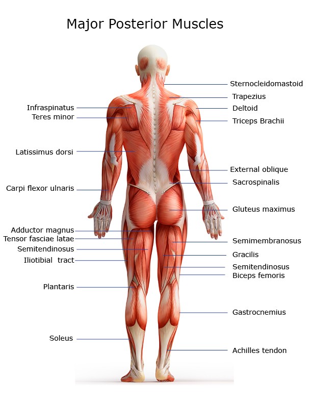
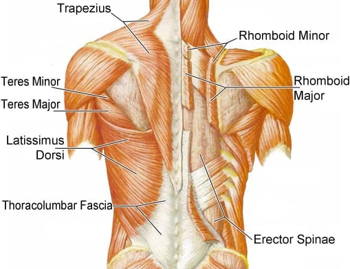
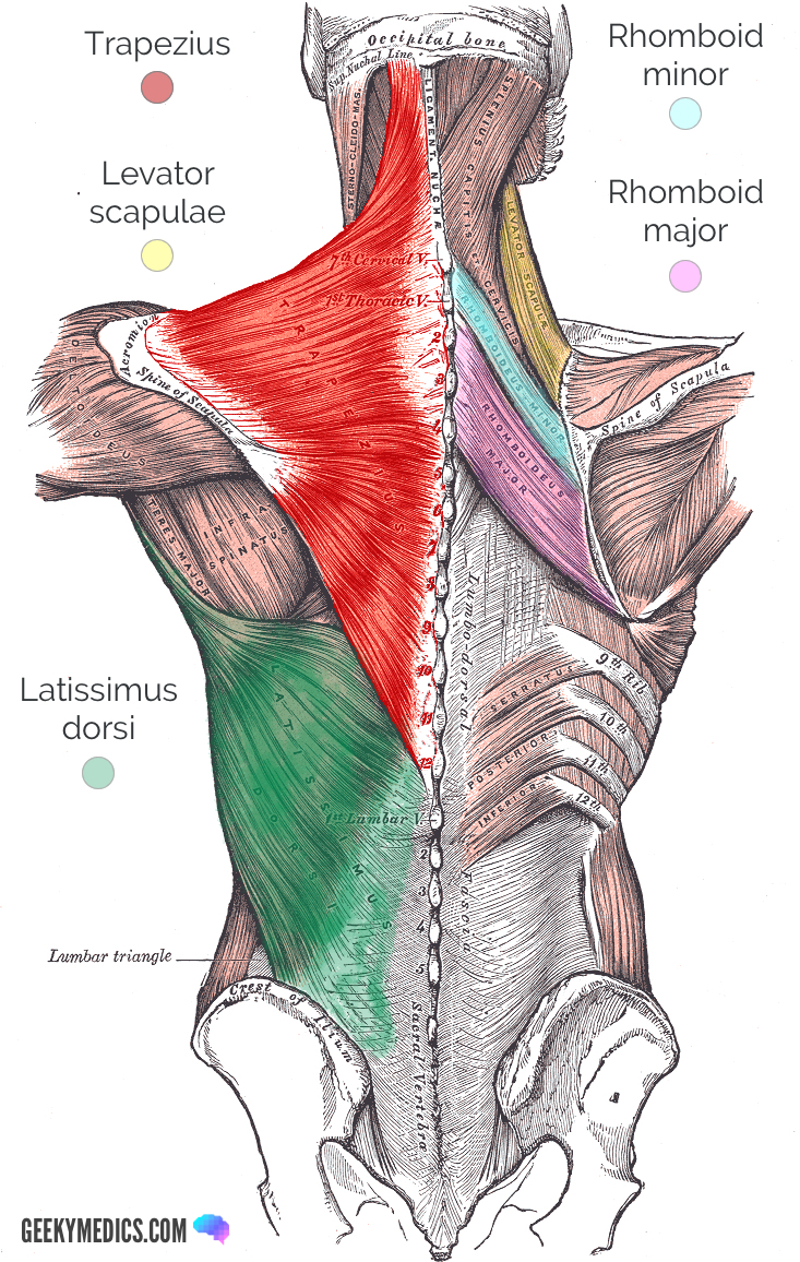
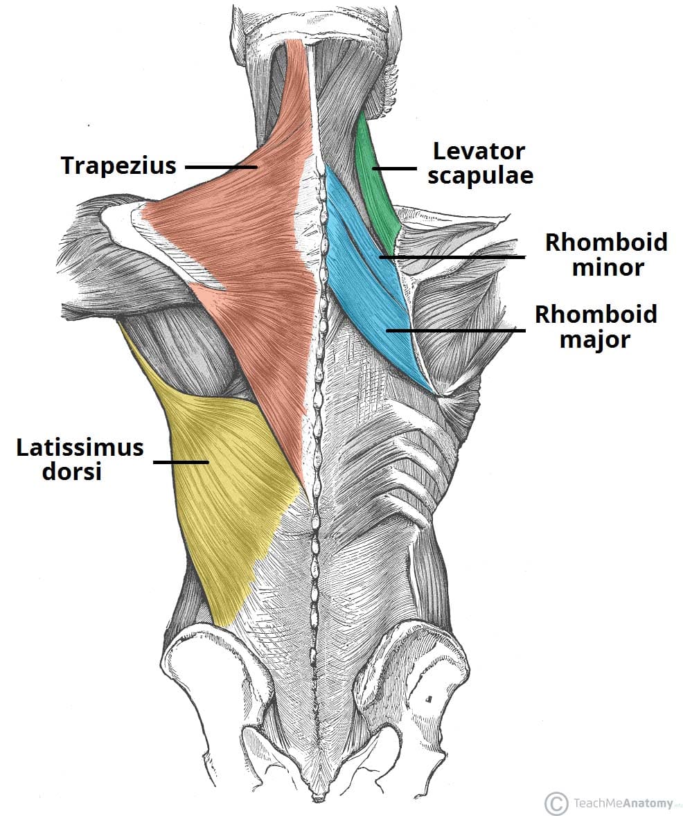
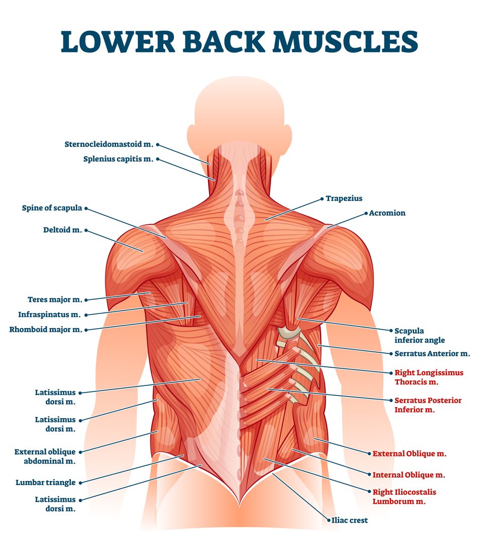
Muscles Of Lower Back Diagram. In this image, you will find an occipital bone, sternocleidomastoid, trapezius, deltoid in Muscles of the lower back diagram. As you can see, there are also have a spine of scapula deltoid, triceps brachii, latissimus dorsi. This picture also contains humerus, olecranon process of ulna, deep to tendon and so on. Anatomical diagrams of the spine and back. This human anatomy module is composed of diagrams, illustrations and 3D views of the back, cervical, thoracic and lumbar spinal areas as well as the various vertebrae. It contains the osteology, arthrology and myology of the spine and back. It is particularly interesting for physiotherapists ... The back anatomy includes the latissimus dorsi, trapezius, erector spinae, rhomboid, and the teres major. The following diagram below is the diagrams of back muscle. On these diagrams of back muscle, you'll learn about back muscles, their locations and functional anatomy. The latissimus dorsi, also known as the "lats" or "wings," are ... For example, some muscles located in the chest also help move the shoulders. Likewise, there are muscles in other parts of the body that help support and move the spine. Below you'll see diagrams along with the names of the back muscles that may be the cause of your pain. How Many Muscles Are in the Back? The back has a total of 40 muscles.
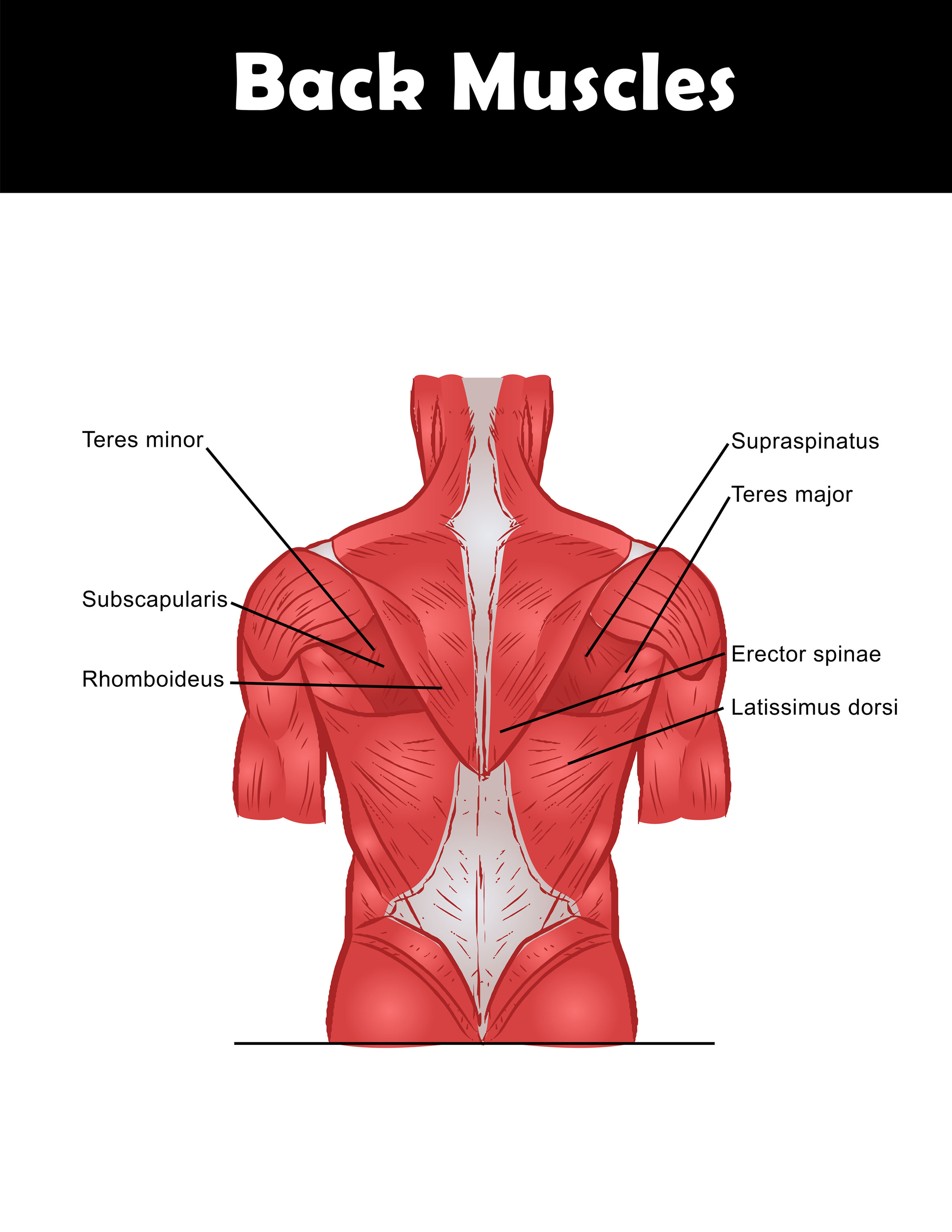
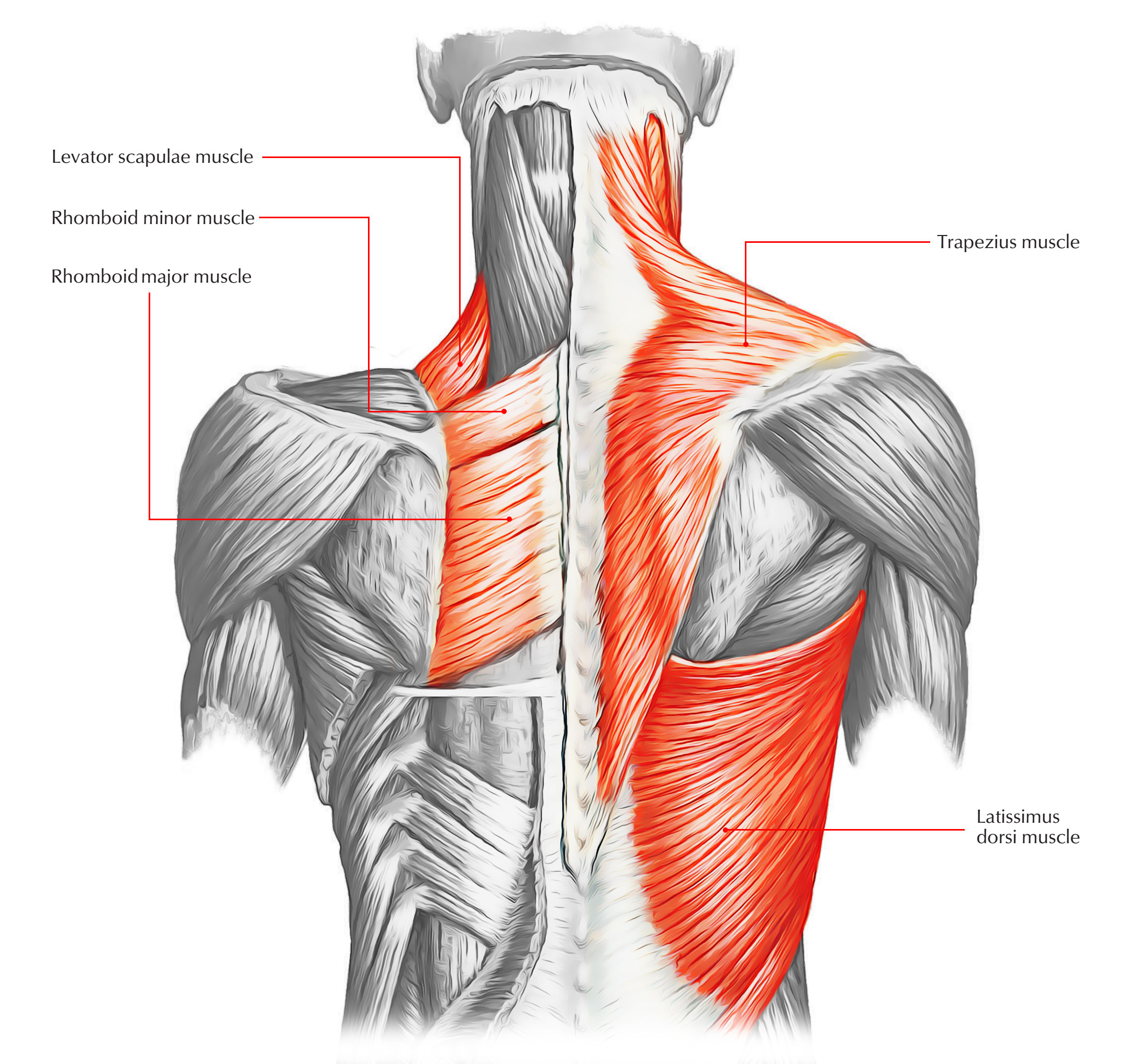
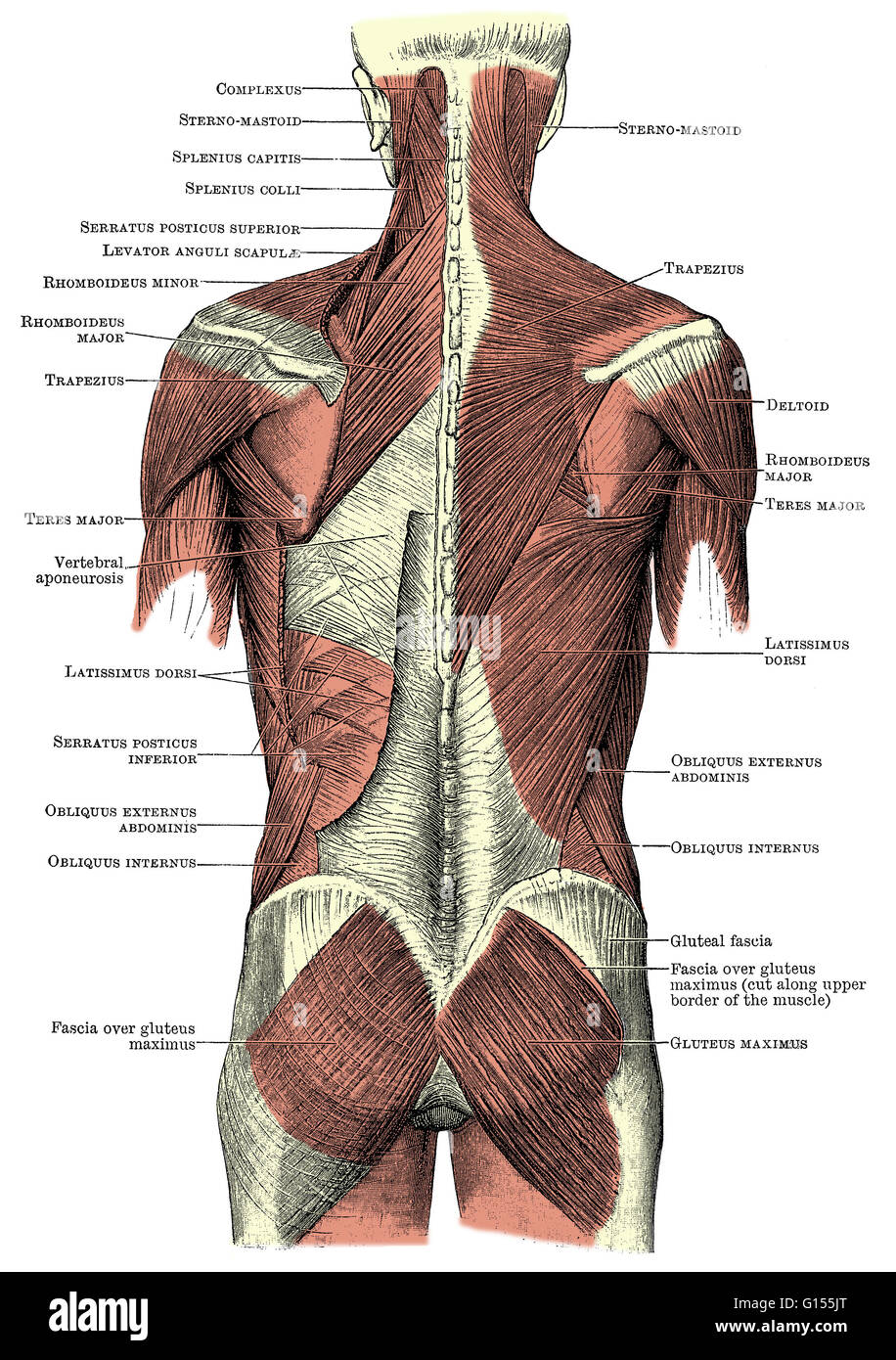
Back muscles. The muscles of the back are a group of strong, paired muscles that lie on the posterior aspect of the trunk They provide movements of the spine, stability to the trunk, as well as the coordination between the movements of the limbs and The back muscles are divided into two large groups: The extrinsic (superficial) back muscles, which lie most superficially on the back. The muscles of the lower back help stabilize, rotate, flex, and extend the spinal column, which is a bony tower of 24 vertebrae that gives the body structure and houses the spinal cord.The spinal ... Muscle layout and location shown in different colours/colors. 3d illustration. diagram of back muscles stock pictures, royalty-free photos & images. Human hand in front and back side. Hand in front and back side on isolated. Illustration about Human body part. diagram of back muscles stock illustrations. The back muscles enable you to stand up straight; support and protect your spine; and reach, pull and extend your arms and torso. Poorly developed back muscles lead to everything from muscle tweaks and pulls to imbalances of the musculature to the all-too-common hunched-over look (the "Neanderthal look").
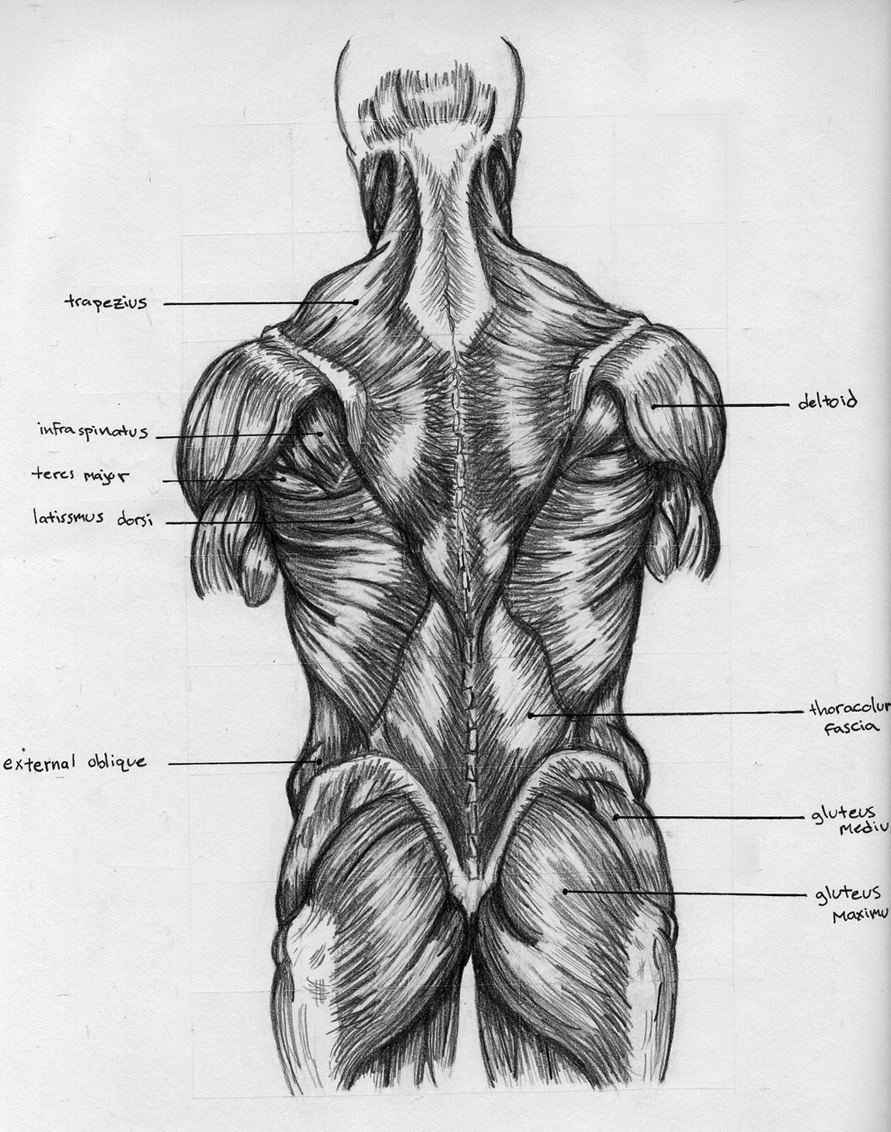
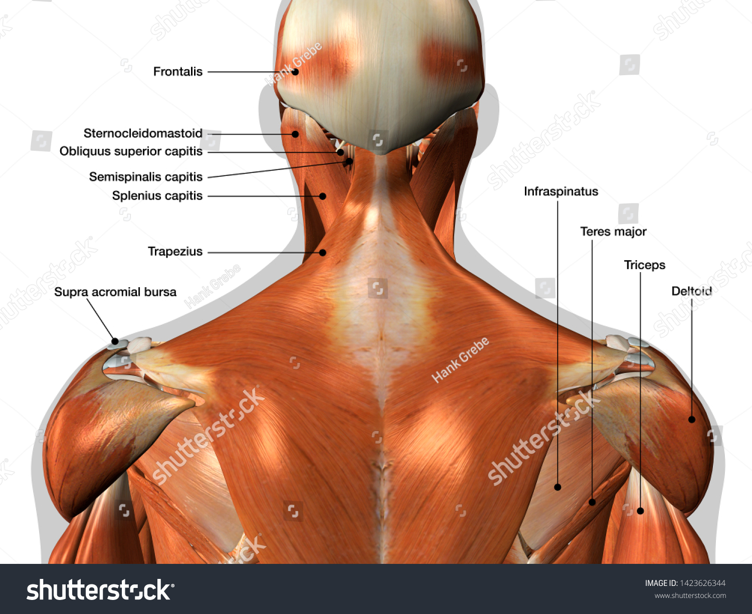
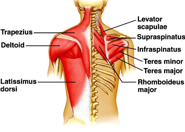
The deltoid, teres major, teres minor, infraspinatus, supraspinatus (not shown) and subscapularis muscles (not shown) all extend from the scapula to the humerus and act on the shoulder joint. The trapezius and latissimus dorsi muscles connect the upper limb to the vertebral column. Both the deltoid and the trapezius are firmly attached to the spine of the scapula.

Anatomy Of The Spine And Back Spine Muscles Diagram. In this image, you will find 1st cervical vertebrae, atlus, cervical plexus, 7th cervical vertebrae, 1st thoracic vertebrae, brachial plexus, spinal dura mater, filaments of spinal nerve roots, 12th thoracic vertebra, 1st lumber vertebra, iliohypogastric nerve, ilioinguinal nerve, lumbar ...
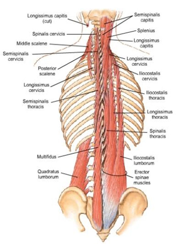
Muscles found in the deep group include the spinotransversales, erector spinae (composed of the iliocostalis, longissimus, and spinalis), the transversospinales, and the segmental muscles. "The best way to strengthen back muscles is in a static position. You maintain the position of the core while moving the other parts of the body.".

The muscles of your back support your spine, attach your pelvis and shoulders to your trunk, and provide mobility and stability to your trunk and spine. The anatomy of your back muscles can be complex. There are several different layers of muscles in your back that are often pulling in different and various directions.

740 lumbar spine anatomy diagram stock photos, vectors, and illustrations are available royalty-free. See lumbar spine anatomy diagram stock video clips. of 8. spinal vertebrae bone spine vertebra toracica spinal cord spine structure back diagram spine sections spinal cord vertebrae spinal structure health diagram. Try these curated collections.


0 Response to "39 diagram of the back muscles"
Post a Comment