38 drag the labels to their appropriate locations on the cycle diagram below.
Drag the labels to their appropriate locations on the diagram of the angiosperm life cycle. Labels can be used once or more than once. Use only white labels for white targets, pink labels for pink targets, and blue labels for blue targets. True False You did not open hints for this part.
Match each of the labels to its correct structure on the diagram Protect your personal email address from spam, bots, phishing and other online abuse. Temporary anonymous email address - no commitments and no risks.
The cycle diagram below shows the sequence of events that affect Ca2+ levels in a muscle cell, beginning with the propagation of an action potential down a T tubule (top of the diagram). Drag the labels to their appropriate locations on the cycle diagram below. Note that SR stands for sarcoplasmic reticulum.
Drag the labels to their appropriate locations on the cycle diagram below.
Drag the appropriate labels to their respective targets. (left from top to bottom) s, g1, cytokinesis (right from top to bottom) interphase, g2, mitosis, mitotic phase 14. During which phase of the cell cycle does DNA duplication, or replication, take place? Interphase When a double helix of DNA is replicated, two complete helices are formed.
Drag the labels to their appropriate locations on the diagram of the water molecules below. Label the following diagram of water molecules indicating the location of bonds and the partial charges on the atoms. Part i of this document describes the smart way to do it. These diagrams tell us that the f 2 molecule has a single bond the co 2 ...
This lets us find the most appropriate writer for any type of assignment. Read more. 100% money-back guarantee. With our money back guarantee, our customers have the right to request and get a refund at any stage of their order in case something goes wrong. Feel safe whenever you are placing an order with us. Read more ...
Drag the labels to their appropriate locations on the cycle diagram below..
Drag each label to the correct location on the graph. Not all labels will be used. Tobias has a job waiting tables where he earns a certain amount every month and an additional amount per table, according to the number of tables he serves. Complete the graph with the appropriate quantities that describe Tobias's earnings over the course of one ...
Drag the labels from the left to their correct locations in the concept map on the right. Part B - Interactions among chromosomes This diagram shows a diploid nucleus (2 n =8) in which chromosome replication has occurred in preparation for mitosis (top right) and meiosis (bottom right). Drag the labels to their appropriate targets to correctly ...
The cycle diagram below shows the sequence of events that affect Ca2+ levels in a muscle cell, beginning with the propagation of an action potential down a T tubule (top of the diagram). Drag the labels to their appropriate locations on the cycle diagram below. Note that SR stands for sarcoplasmic reticulum. • Show less
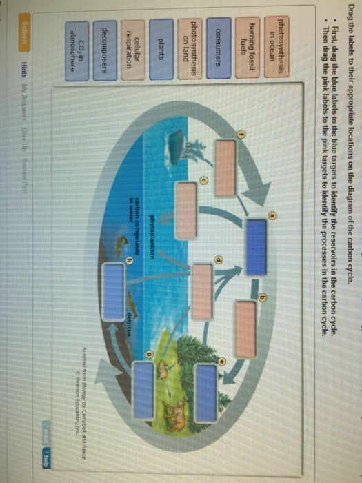
Drag the labels to their appropriate locations on the diagram of the carbon cycle. First, drag the blue labels to the blue targets to identify the reservoirs in the carbon cycle. Then drag the pink labels to the pink targets to identify the processes in the carbon cycle.
The cycle diagram below shows the sequence of events that affect Ca2+ levels in a muscle cell, beginning with the propagation of an action potential down a T tubule (top of the diagram). Drag the labels to their appropriate locations on the cycle diagram below. Note that SR stands for sarcoplasmic reticulum.
11.12.2008 · Abstract. Web Content Accessibility Guidelines (WCAG) 2.0 covers a wide range of recommendations for making Web content more accessible. Following these guidelines will make content accessible to a wider range of people with disabilities, including blindness and low vision, deafness and hearing loss, learning disabilities, cognitive limitations, limited movement, …
Drag the correct label to the appropriate location in the table. Labels can be used once, more than once, or not at all. Predicted eye color(s): all wild type all wild type all wild type 1/2 wild type 1/2 vermilion. Red-green color blindness is due to an X-linked recessive allele in humans.
Academia.edu is a platform for academics to share research papers.
Jun 04, 2021 · Label Heart Anatomy Diagram Printout – EnchantedLearning. Hover over the centre of the text and drag to reposition the text, or hover over the top rotation handle and drag to rotate the text. Feb 01, 2017 · Drag the labels to their appropriate locations to complete the Punnett square for Morgan’s F 1 x F 1. Drag shapes from ...
Drag the labels to their appropriate locations on the diagram of the carbon cycle. Reservoirs are colored blue, and processes of the carbon cycle are in pink.-CO2 in the atmosphere-Photosynthesis on land-Plants-Consumers-Decomposers-Cellular respiration-Photosynthesis in ocean-Burning fossil fuels
© 2021 Houghton Mifflin Harcourt. All rights reserved. Terms of Purchase Privacy Policy Site Map Trademark Credits Permissions Request Privacy Policy Site Map ...
The table below the diagram confirms this and tells us the significance values of the three comparisons. The significance value of the comparison between Friends and EastEnders is 0.037, which is below the criterion of 0.05, therefore we can conclude that EastEnders led to significantly more arguments than Friends.
Drag the labels to their appropriate locations in the figure. First, drag labels to targets (a) and (b) to indicate whether these environments are hydrophilic or hydrophobic. Next, drag the phospholipid layers to targets (c) and (d) to indicate how they are oriented in the plasma membrane.
Recall that you can open the Workflow diagram by right-clicking ToyCatapult.aird in the Capella Project Explorer and clicking Open Activity Explorer. Click the Systems Analysis chevron, or its corresponding tab at the bottom of the window. Locate and click the Contextually create new System Actors from Operational Entities/Actors button. In the resulting dialog, (1) select all of …
Drag the labels to their appropriate locations on the diagram below. Targets of Group 1 can be used more than once. a. hydrogen bond. The DNA double helix is composed of two strands of DNA; each strand is a polymer of DNA nucleotides. Each nucleotide consists of a sugar, a phosphate group, and one of four nitrogenous bases. The structure and orientation of the two …
Transcribed image text: Drag the labels to their appropriate locations on the cycle diagram below. Note that SR stands for sarcoplasmic reticulum. cytosolic Ca2+ level rises Ca2+ diffuses out of myofibril cytosolic Ca2+ level drops Ca2+ diffuses into myofibril Ca2+ diffuses out of SR Ca2+ pumped into SR action potential propagates down t tubule Ca2+ channels in SR open Ca2+ channels in SR ...
Part A - Hydrogen bonding Label the following diagram of water molecules, indicating the location of bonds and the partial charges on the atoms. Drag the labels to their appropriate locations on the diagram of the water molecules below. Labels can be used once, more than once, or not at all. Hint 1.
Identify the structures and determine which hypha is septate and which is coenocytic. (Note that although this diagram shows the two types of hyphae, a fungus can have either one type or the other, but not both.) Drag the labels to their appropriate locations on the diagram of the fungus and hyphae below.
The diagram below illustrates the life cycle of Dictyostelium, a cellular slime mold. Label the diagram to indicate the processes that are occurring as well as whether each cell type is haploid or diploid. Drag the labels to their appropriate locations on the diagram. Use the pink labels for pink targets.
Drag the labels to their appropriate locations on the diagram of the water molecules below. Label the following diagram of water molecules. When two water molecules are in the vicinity of one another the slightly negative oxygen atom of one molecule is. Explain what a hydrogen bond is as compared to covalent and ionic bonds.
Drag the labels to their appropriate locations on the cycle. Drag the labels to their appropriate locations on the cycle diagram below. Note that SR stands for s…. Show more Drag the labels to their appropriate locations on the cycle diagram below. Note that SR stands for sarcoplasmic reticulum. • Show less.
Drag the labels to their appropriate locations on the diagram of the water molecules below. Label the following diagram of water molecules indicating the location of bonds and the partial charges on the atoms. There are two lone pairs of electrons on each oxygen atom represented by. Labels can be used once more than once or not at all.
Drag the labels to their appropriate locations on the diagram below. Pink labels can be used more than once. [The DNA double helix is constructed from two strands of DNA, each with a sugar-phosphate backbone and nitrogenous bases that form hydrogen bonds, holding the two strands together. Each DNA strand has two unique ends.
Drag and drop the text labels onto the boxes next to the heart diagram. The respiratory system objectives after this lesson students will be able to d411. Write each step of the process in a separate box in the order in which it occurs. Drag each label to the appropriate location on this diagram of the human respiratory system.
drag the labels to their appropriate locations on the diagram of the neurons below. use targets of group 1 to indicate the components of neurons. use targets of group 2 to indicate the locations of gated ion channels.
The cycle diagram below shows the sequence of events that affect Ca2+ levels in a muscle cell, beginning with the propagation of an action potential down a T tubule (top of the diagram). Drag the labels to their appropriate locations on the cycle diagram below. Note that SR stands for sarcoplasmic reticulum.
The cycle diagram below shows the sequence of events that affect Ca2+ levels in a muscle cell, beginning with the propagation of an action potential down a T tubule (top of the diagram). Drag the labels to their appropriate locations on the cycle diagram below. Note that SR stands for sarcoplasmic reticulum.
The diagram below shows a replication fork with the two parental DNA strands labeled at their 3' and 5 4/4 (5). The diagram below shows a bacterial replication fork and its principal proteins. Drag the labels to their appropriate locations in the diagram to describe the name or function of each structure. Use pink labels for the pink targets ...
Use this diagram to track the carbon-containing compounds that play a role in these two stages. Drag the labels from the left (which represent numbers of carbon atoms) onto the diagram to identify the number of carbon atoms in each intermediate in acetyl CoA formation and the citric acid cycle. Labels may be used more than once.
Follow these steps 1. First, drag blue labels onto blue targets only to identify each stage of the life cycle 2. Next, drag pink labels onto pink targets only to identify the process by which each stage occurs 3. Then, drag white labels onto white targets only to identify the ploidy level at each stage. Labels can be used once, more than once ...
Drag the labels to their appropriate locations on the diagram of the carbon cycle. First, drag the blue labels to the blue targets to identify the reservoirs in the carbon cycle. Then drag the pink labels to the pink targets to identify the processes in the carbon cycle. Submit Hints My Answers Give Up Review Part.

0 Response to "38 drag the labels to their appropriate locations on the cycle diagram below."
Post a Comment