42 cow eye dissection diagram
the white of the eye 80 The intrinsic eye muscles are under the control of which of the following? (Circle the correct response.) autonomic nervous system Dissection of the Cow (Sheep) Eye somatic nervous system 90 What modification of the choroid that is not present in humans is found in the cow eye? COW no What is its function? c 10. COW EYE DISSECTION · 1. Examine the outside of the eye. · 2. Locate the covering over the front of the eye, the cornea. · 2. Cut away the fat and muscle, this may ...
Cow Eye Dissection. STUDY. Learn. Flashcards. Write. Spell. Test. PLAY. Match. Gravity. Created by. Ms_Finn TEACHER. Terms in this set (26) Iris. A ring of muscle tissue that forms the colored portion of the eye around the pupil and controls the size of the pupil opening. Lens. The transparent structure behind the pupil that changes shape to ...
Cow eye dissection diagram
About Press Copyright Contact us Creators Advertise Developers Terms Privacy Policy & Safety How YouTube works Test new features Press Copyright Contact us Creators ... Eye Dissection Diagram. Module 1 Labeled Diagram Of The Eye Diagram Of The Eye Dot Worksheets Diagram. Cow Eye Dissection Worksheet Cow Eyes Dissection Frog Dissection Worksheet. Diagram Of Basic Parts Of The Eye Eye Anatomy Parts Of The Eye Diagram Of The Eye. Eyes Layers Of Learning Human Eye Diagram Parts Of The Eye Human Eye. Central Retinal Artery - The central retinal artery supplies blood to the retina as it branches into smaller segments upon leaving the optic disc. Central retina vein - the vessel that carries blood away from the retina. Choroid - the thin, blood-rich membrane that lies between the retina and the sclera; responsible for supplying blood to the retina.
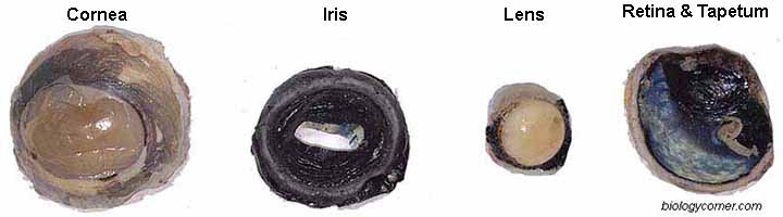
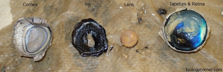
Cow eye dissection diagram. The iris is a muscle that controls how much light goes into the eye and suspended between the cornea and lens. As we have seen in the dissection a cow's iris is brown. The pupil is the dark circle that's in the center of the iris and it lets light into the inner eye. DISSECTION OF THE COW EYE Please make sure to wear gloves and safety glasses when you are dissecting, and make sure to clean up thoroughly after the lab. Also, the cow eyes can be rather slippery, so use caution when handling and cutting them. You will need a scalpel and forceps. 1. First, identify the most external structures of the eye. 1 day ago · Featured craft: "White as Snow" Globe. Jun 18, 2021 · Download MCQs Questions with Answers of Classes 12, 11, 10, 9, 8, 7, 6, 5, 4, 3, 2, 1 through quick links available. COW’S EYE DISSECTION – good virtual dissection with no cost or mess to clean up afterwards. Try this to find out. Do a cow eye dissection. And get our free printable eye diagram to label and color. The Human Eye Labeling Activity (teacher made). This KS2 The Human Eye Labelling Activity supports the teaching of the anatomy of the eye and visual process. Cow Eye Dissection. Teacher's guide for dissecting a cow or a sheep eye.
cow eye, dissection Remaining 0. Correct 0. Wrong 0. Press play! 0%. 0:00.0. Quit. Again. This game is part of a tournament. You need to be a group member to play the tournament. Join group, and play Just play. Your Scorecard. The scorecard of a champion. Score . 0 % Time . 0:00.0. Place # 0. Learn how to dissect a cow's eye in your classroom. This resource includes: a step-by-step, hints and tips, a cow eye primer, and a glossary of terms. Cow's Eye Dissection - Eye diagram This is a great activity for middle and high school students. It allows them to participate in a dissection, while teaching the anatomy and structure of the eye. To get started, watch the YouTube video Cow's Eye Dissection: Exploratorium. It can be a support for guiding instruction. It offers good explanations in age appropriate language. You need to enable JavaScript to run this app. Kahoot! You need to enable JavaScript to run this app.
Cow eye dissection lab structure and function of the human eye cow eye dissection kids health topics eyes how your structure and function of the eyes. Part 2 The Cow Eye Bctc. A P Lab Unit 2 Labeled Cow Eye Diagram Quizlet. Cow Eye Dissection Diagram Quizlet. Eye Anatomy. Kids Health Topics Eyes How Your Work. 19 Best Cow Eye Dissection Labeled. Jul 30, 2014 - Cow Eye Dissection Diagram Labeled | Cow Eye Review the glossary provided at the end of this dissection guide. Refer to the diagram of the eye as a general reference as you observe and identify external and internal structures. 1. Observe the outer structure of the eye. Identify the following: optic nerve, sclera, cornea. 2. Trim away excess tissue surrounding the eyeball on the sclera. 3. Cow Eye Dissection: Directions and questions for dissecting a cow eye. Also includes a diagram of the eye, fun facts, an activity on how to find your blind spot and all answer keys. Appropriate for grades 4-7, I run this lab for each of the 4th grade classes at my school. It's a HUGE hit!!! This
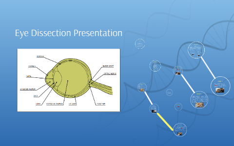
Eye Dissection Presentation By Joshua Lee
This is an online quiz called Cow Eye Labeling. There is a printable worksheet available for download here so you can take the quiz with pen and paper. Your Skills & Rank. Total Points. 0. Get started! Today's Rank--0. Today 's Points. One of us! Game Points. 11. You need to get 100% to score the 11 points available.
2
The Cow Eye Dissection Lab. What are the structures of the mammalian eye and how do they function? The mammalian eye consists of many specialized cells and ...
Solved Please Label The Diagram Of Cow S Eye Dissection Chegg Com
Start studying Cow Eye Dissection & Parts of the Eye. Learn vocabulary, terms, and more with flashcards, games, and other study tools.

Elementary Dissection Mat Carolina Cow Eye Carolina Com
Study mammalian organ anatomy as you dissect the four preserved specimens in this organ dissection kit: a cow eye, a sheep heart, a sheep brain, and a sheep kidney.. Providing an interactive biology lab or life science activity for middle school and high school students, this complete organ dissection kit includes: full-color photographic dissection guides for all four specimens,

Biology Lab 10 Cow Eye Dissection Diagram Quizlet
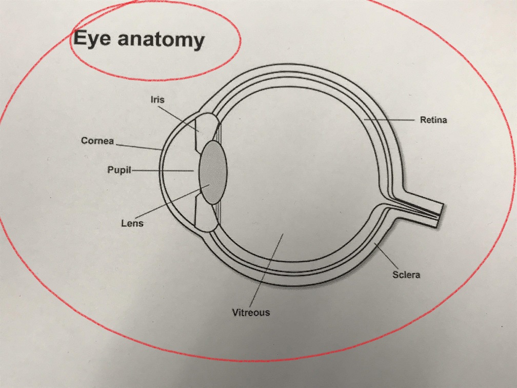
Cow Eye Dissection (At Home) Use the following diagram and fill in the diagram as you visually examine the eye. 1. Examine the outside of the eye. See how many parts of the eye you can identify. You should be able to find the whites (or sclera), the tough, outer covering of the eyeball. You should also be able to identify the fat and muscle surrounding the eye.
Ppt Cow Eye Dissection Powerpoint Presentation Free Download Id 3482425
Cow Eye Dissection: Examining Structure and Function. Carolina Biological Supply Company 2 Extension Activities ... Use the diagram to identify the internal structures of the eye. 6. Remove the vitreous humor and lens from the front portion of the eye. a. Describe the vitreous humor. Why must the vitreous humor

Cow Eye Dissection Free Essay Example
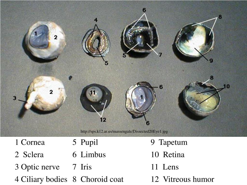
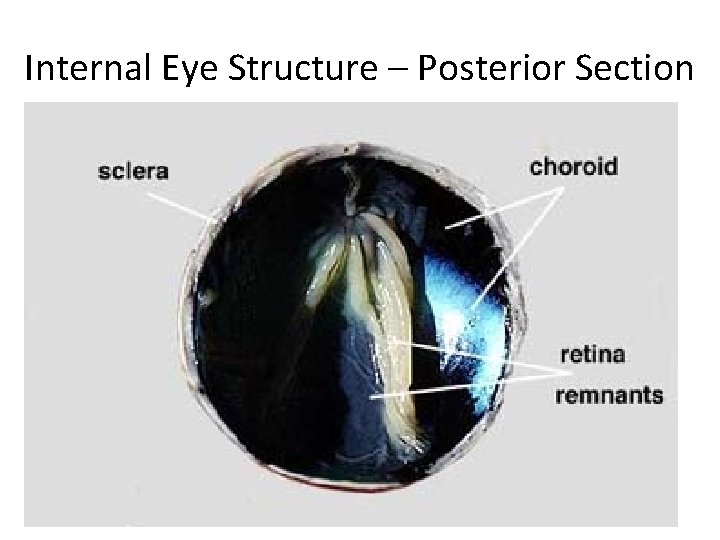
Dissection 101: Cow Eye. Use the point of a scissors or a scalpel to make an incision through the layers of the eye capsule (similar to figure 1); there are three layers from the exterior: sclera, whitish/grey, continuous with the transparent cornea, choroid, thin dark black layer and the retina, thin greyish/pink layer.
Dissection Cattle Anatomy Human Eye Png Clipart Anatomy Biology Blue Glow Cattle Diagram Free Png Download
Learn about cow stomachs. Look at different types of cows. (*)Just for fun if you like: If you only have a first or second grader, you could skip this. You can go here and choose a craft or coloring page. You could also read this fable. Level 5-8. Learn about cow stomachs. Look at the different types of cows and learn about them. Save the cows ...
2
YOUR GUIDE TO DISSECTION Since 1817 Dissecting an Eye Before dissection, allow the students to a have a good look at the Eye and see if they can identify any parts of the Eye. Before commencing any kind of dissection on animal material, always read and implement any Health & Safety measures.

Parts Of The Eye Cow Eyes Anatomy And Physiology Eye Anatomy
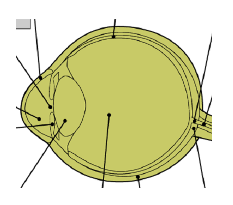
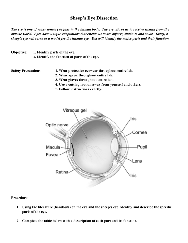
If you scroll back up, you will see a diagram of the cows eye as well as a table of the parts and functions. 4. Which method, a diagram, model or dissection helped you to understand the parts of the eye the most? Why? The method that helped me learn the parts of the eye best was the diagram as well as the dissection lab.

Anatomy Of Cow Eye Anatomy Drawing Diagram
A diagram of ultrasonic signals emitted by a bat, and the echo from a nearby object As in all other tetrapods, mammals have a larynx that can quickly open and close to produce sounds, and a supralaryngeal vocal tract which filters this sound.

Sheep Eye Dissection Virtual Practical Exam Practice Quiz For Anatomy Youtube
Cow eye, dissecting pan, dissecting kit, safety glasses, lab apron, and gloves. Procedure (External Structure): Obtain a cow eye, place it in your dissecting pan, & rinse the eye with water. Rotate the eye until the larger bulge or tear gland is on the top of the eye. The eye is now in the position it would be in a body as you face the body. On ...
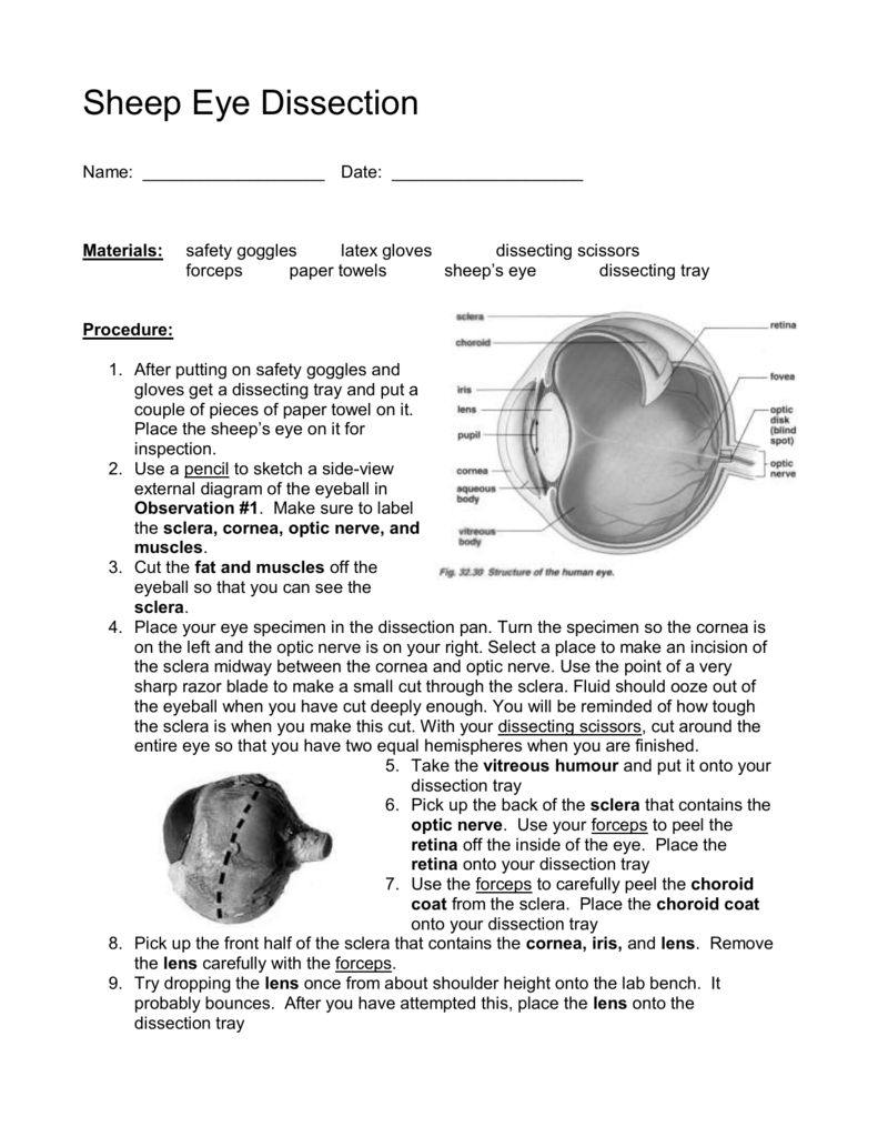
Sheep Eye Dissection Lab
SHEEP EYE DISSECTION PROCEDURES The anatomy of the human eye can be better shown and understood by the actual dissection of an eye. One eye of choice for dissection, that closely resembles the human eye, is that of the sheep. Differences between the two eye types will be mentioned as the dissection is completed.
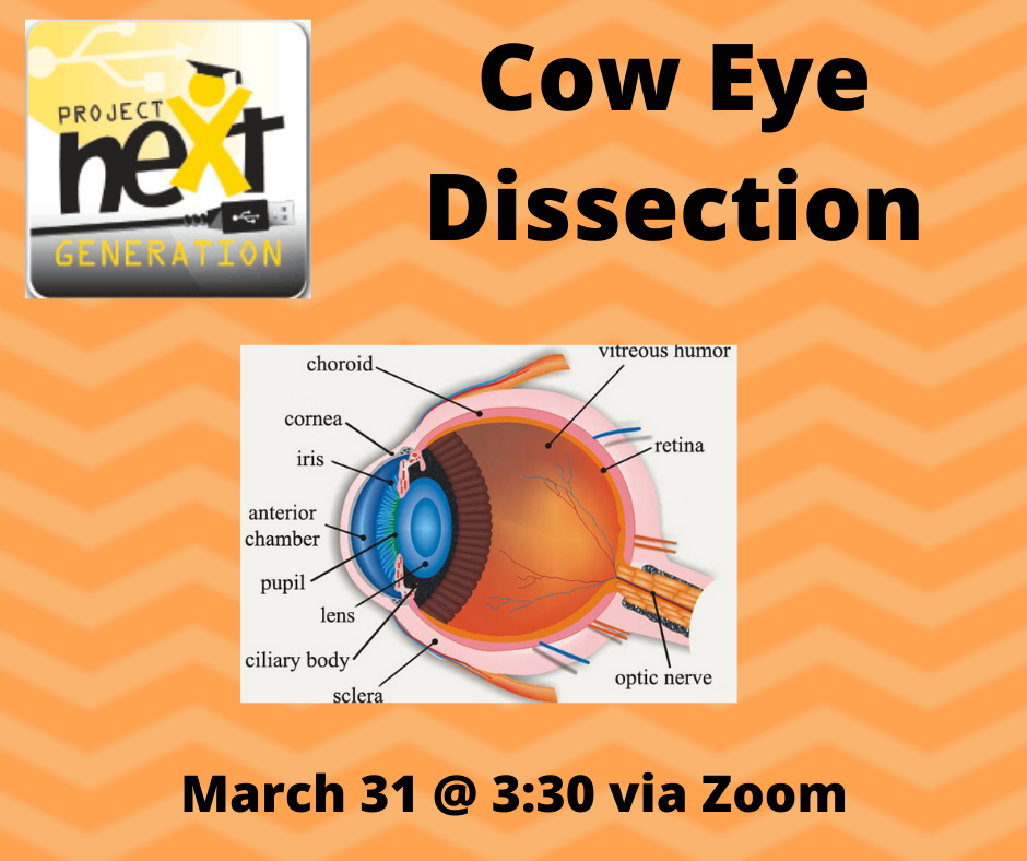
Cow Eye Dissection
At the Exploratorium, we dissect cows’ eyes to show people how an eye works. This Web site shows photos and videos of a dissection. If you try this at home, wash your hands after the dissection. Wear latex gloves if you have cuts in your hands. Here’s a cow’s eye from the meat company.
Cow Eye
Cow Eye Dissection: Directions and questions for dissecting a cow eye. Also includes a diagram of the eye, fun facts, an activity on how to find your blind spot and all answer keys. Appropriate for grades 4-7, I run this lab for each of the 4th grade classes at my school.
Sheep Eye Dissection Hrsbstaff Ednet Ns Caholmesdl Virtualsheep
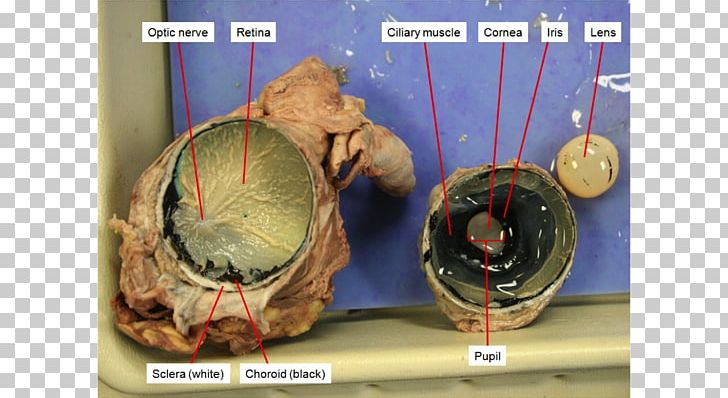
Cow Eye Dissection: Internal Anatomy. 1. Place the cow eye on a dissecting tray. The eye most likely has a thick covering of fat and muscle tissue. Carefully cut away the fat and the muscle. As you get closer to the actual eyeball, you may notice muscles that are attached directly to the sclera and along the optic nerve.
2
Cow Eye Dissection. The mammalian eye is a sensory organ that operates as part of the nervous system. These complex organs gather light, focus it on receptor cells, and transmit the information to the brain where it is interpreted. Placement and shape of eyes vary across the animal kingdom, but the main function remains consistent—vision.

Eye Dissection
BIOL 145 Human Anatomy and Physiology Dissection* (1 Hour) Prerequisites : BIOL 144 and department approval. Students will dissect the cat and study the relationship of structures to function in the organ systems of the cat. In this laboratory course, they will also dissect the cow kidney, heart, brain and eye.
Cow Eye Dissection Welcome To Cow Eye Dissection This Powerpoint Was Created To Be Downloaded And Used With Your Students Please Feel Free To Edit Ppt Download
COW'S EYE dissection page 6 Now take a look at the rest of the eye. If the vitreous humor is still in the eyeball, empty it out. On the inside of the back half of the eyeball, you can see some blood

Funventure Cow Eye Dissection Discovery Outpost Medicine Park October 1 2021 Allevents In
• Preserved cow's eye • Forceps • Dissection probes • Dissection scissors • Dissection tray • Vinyl or latex gloves • Apron or tee-shirt • Paper towels • Plastic trash bag External features of the eye A. Locate the cornea, sclera, and optic nerve. a. The white part of the eye, the sclera, is a tough, outer covering
1
Central Retinal Artery - The central retinal artery supplies blood to the retina as it branches into smaller segments upon leaving the optic disc. Central retina vein - the vessel that carries blood away from the retina. Choroid - the thin, blood-rich membrane that lies between the retina and the sclera; responsible for supplying blood to the retina.
Png Cow Eye Dissection Moline Public Library
Eye Dissection Diagram. Module 1 Labeled Diagram Of The Eye Diagram Of The Eye Dot Worksheets Diagram. Cow Eye Dissection Worksheet Cow Eyes Dissection Frog Dissection Worksheet. Diagram Of Basic Parts Of The Eye Eye Anatomy Parts Of The Eye Diagram Of The Eye. Eyes Layers Of Learning Human Eye Diagram Parts Of The Eye Human Eye.

Cow Eye Dissection Guide Google Slides
About Press Copyright Contact us Creators Advertise Developers Terms Privacy Policy & Safety How YouTube works Test new features Press Copyright Contact us Creators ...
Hamburg Csd 5th Grade Cow Eyes

What Are The Parts Of The Eye Ppt Download
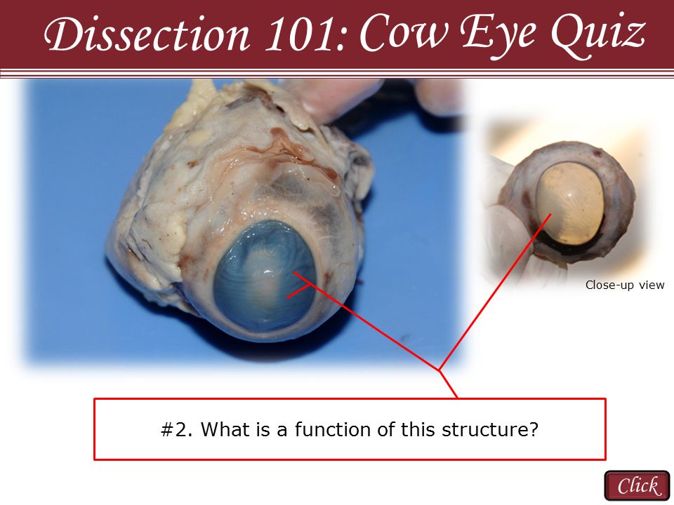
Cow Eye Quiz Dissection 101 Click Ppt Video Online Download
Sheep Eye Dissection

Cow Eye Dissection Perkins Elearning

Use The Pictures Below To Name The Parts Of The Eye That You Will Observe In Course Hero
2
Cow Eye Dissection

Cow Eye Dissection Diagram Quizlet

Activities And Answer Keys Ck 12 Foundation

Cow Eye Dissection Carolina Com
Human

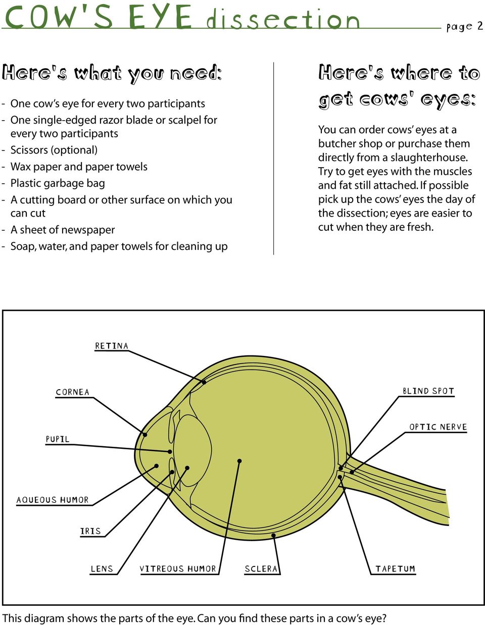
Cow S Eye Dissection Eye Diagram
Human
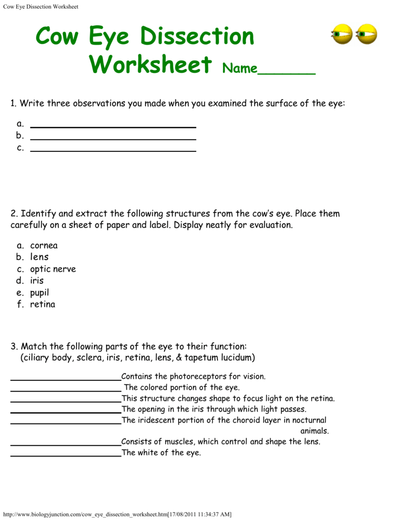
Cow Eye Dissection Worksheet
2
Cow S Eye Dissection Dissecting A Cow S Eye Step By Step Instructions Safety First Pdf Free Download

Cow Eye Dissection Diagram Part 1 Diagram Quizlet

Cow Eye Dissection Biology4friends
0 Response to "42 cow eye dissection diagram"
Post a Comment