39 on the diagram below identify alveolar epithelium
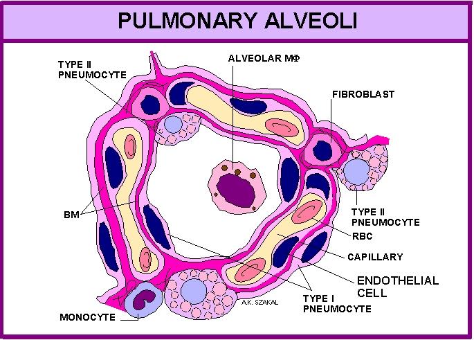
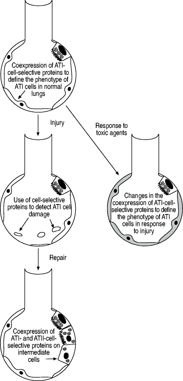
The diagram below contains a diagram of a Type I cell with six cytoplasmic plates and an informative SEM image of isolated Type I cells in culture. That latter picture is from Fuchs et al (2003) and shows them spreading across a plate, forming thin disks with centrally raised blobs containing the nuclei and all the other cellular machinery. Review Sheet 23 Anatomy Respiratory Review Sheet 23 On the diagram below, identify alveolar epithelium, capillary endothelium, alveoli, and red blood cells. Bracket the respiratory membrane. VÉeu É Elastic fiber O st W Connective-tissue fibers I Monocyte Connective-tissue cell Why does oxygen move from the. PDF Review Sheet 23 Anatomy Respiratory System Diagram system diagram is available in ...
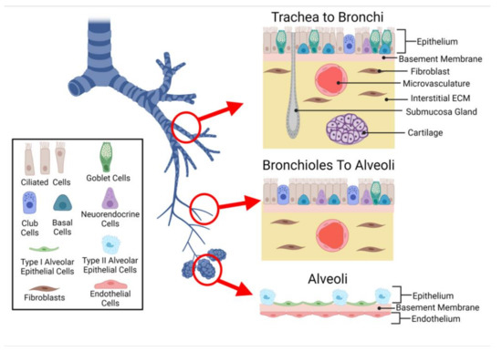
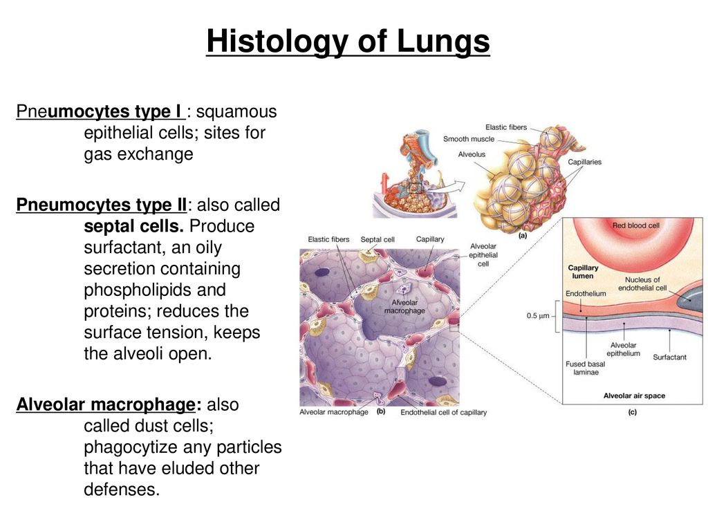
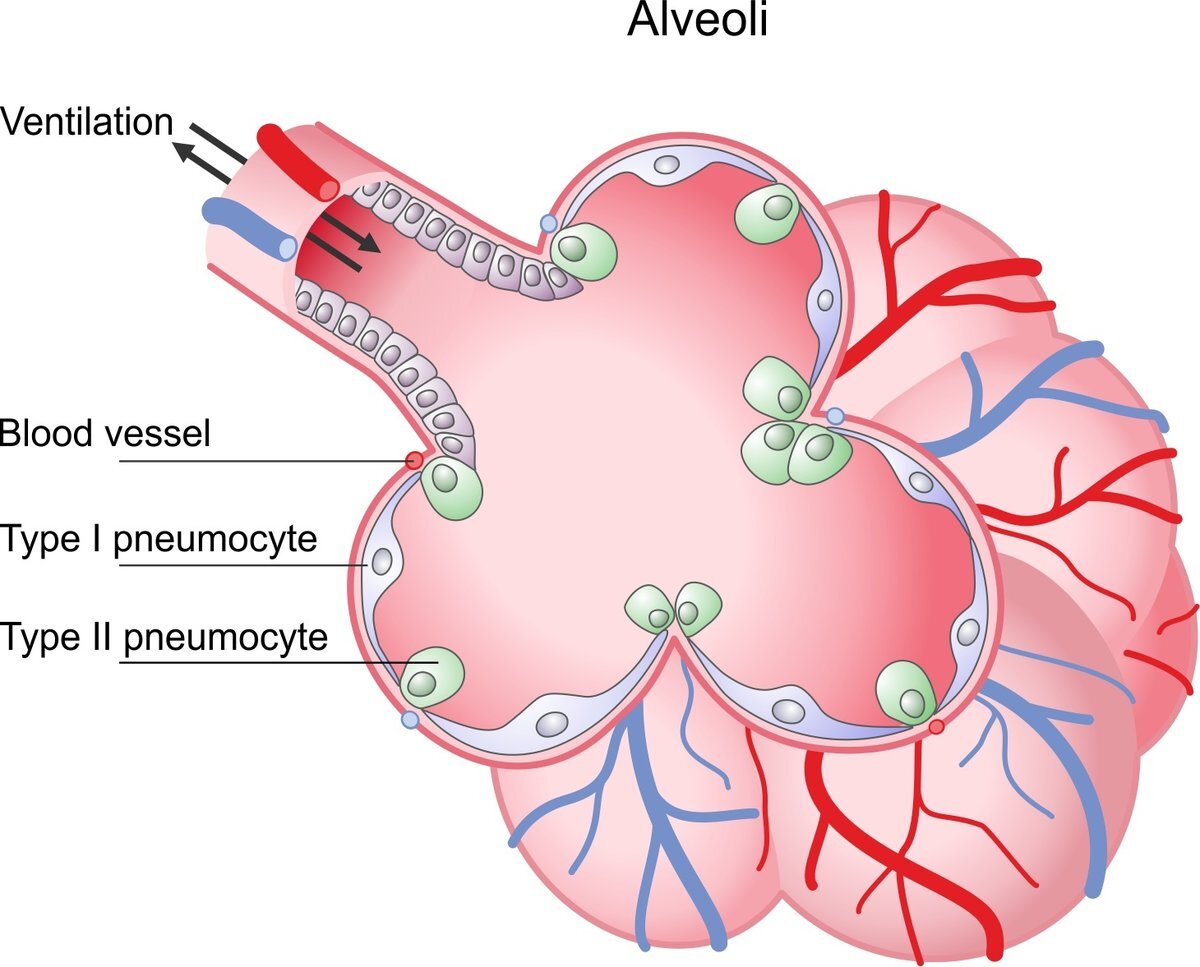
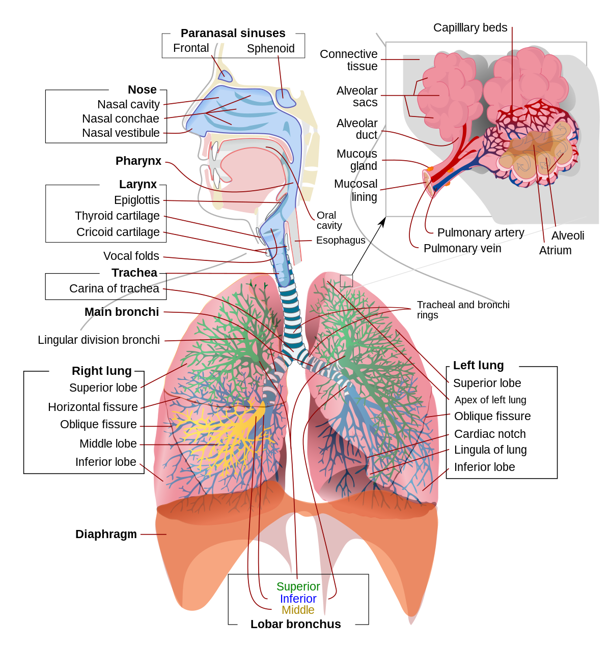
This diagram shows a diagram of an alveolar sac, showing how the organisation of the alveoli, and the network of blood capillaries that surround the alveoli (in red). The epithelium of the alveoli, contains two main types of cells: type I pneumocytes: large flattened cells - (95% of the total alveolar area) which present a very thin diffusion barrier for gases. type II pneumocytes (making up 5 ...
On the diagram below identify alveolar epithelium
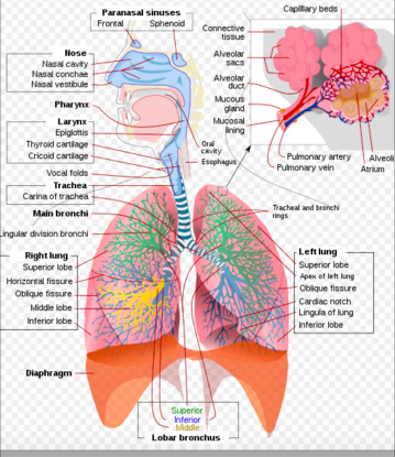
On the diagram below identify alveolar epithelium capillary. Leading Edje Home Bronchials trace a molecule of oxygen from the nostrils to the pulmonary capillaries of the lunges. Appropriately label all structures provided with leader lines on the diagrams below. Back to notecard set easy notecards home page. Review sheet exercise 36. 284 review sheet 36 produce a serous fluid that reduces ... Cecie Starr, Ralph Taggart, Christine Evers · 2012 · ScienceLabeling and Matching [pp.682-683] Identify each structure indicated in the diagram of the respiratory membrane below. ... _____ B. alveolar epithelium 23. Question: iagram below, identify alveolar epithelium, capillary endothelium, alveoli, and red blood cells. Bracket the brane respira Elastic fiber Connective tissue fibers Monocyte . This problem has been solved! See the answer See the answer See the answer done loading. Show transcribed image text Expert Answer . Who are the experts? Experts are tested by Chegg as specialists in their subject ...
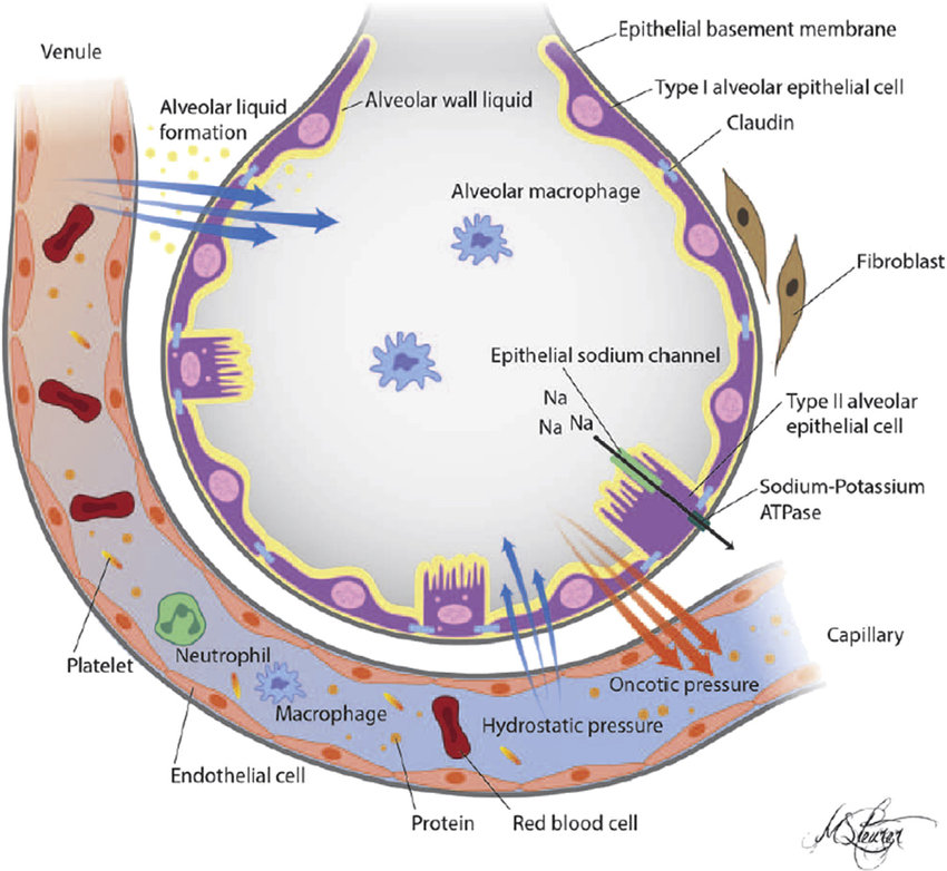
On the diagram below identify alveolar epithelium. Diagram of alveolus and ion channels, pumps, and pores in the alveolar epithelium. The alveolus is composed of alveolar epithelial type 1 cells, alveolar ... Another kind of stratified epithelium is transitional epithelium, so-called because of the gradual changes in the shapes and layering of the cells as the epithelium lining the expanding hollow organ is stretched. Transitional epithelium is found only in the urinary system, specifically the ureters and urinary bladder. When the bladder is empty, this epithelium is convoluted and has cuboidal ... Underneath the thin skin of the nose are its skeletal features. ... An olfactory epithelium used to detect odors is found deeper in the nasal cavity. On the diagram below identify alveolar epithelium capillary. Appropriately label all structures provided with leader lines on the diagrams below. Label all structures provided with leader lines on the diagram below. Fill in the blanks with the terms provided. Plveoll superior lobe middle lobe inferior lobe. Correctly label all structures provided with leader lines in the diagram of a molar ...
On the diagram below identify aveolar epithelium, capillary endothelium, alveoli, land red blood cells. Bracket the respiratory membrane. Elastic.4 pages On the diagram below, identify the alveolar duet, respiratory bronchioles, terminal bronchiole, alveoli, and alveolar sa. Examining Prepared Slides of Tracheal and Lung Tissue 14. The tracheal epithelium is ciliated and has goblet cells. What is the function of each of these modifications? cilia: goblet cells: 15. The tracheal epithelium is said to be pseudostratified. Why? 16. What structural ... Solution for 14. On the diagram below, identify alveolar epithelium, capillary endothelium, alveoli, and red blood cells. Bracket the respiratory membrane.… On the diagram below, identify alveolar epithelium, capillary endothelium, alveoli, and red blood cells. Bracket the respiratory membrane. VÉeu É Elastic fiber O st W Connective-tissue fibers I Monocyte Connective-tissue cell 15. Why does oxygen move from the alveoli into the pulmonary capillary blood? CIS 770 16. What structural characteristic of the alveoli makes them an ideal site for the ...
Question: iagram below, identify alveolar epithelium, capillary endothelium, alveoli, and red blood cells. Bracket the brane respira Elastic fiber Connective tissue fibers Monocyte . This problem has been solved! See the answer See the answer See the answer done loading. Show transcribed image text Expert Answer . Who are the experts? Experts are tested by Chegg as specialists in their subject ... Cecie Starr, Ralph Taggart, Christine Evers · 2012 · ScienceLabeling and Matching [pp.682-683] Identify each structure indicated in the diagram of the respiratory membrane below. ... _____ B. alveolar epithelium 23. On the diagram below identify alveolar epithelium capillary. Leading Edje Home Bronchials trace a molecule of oxygen from the nostrils to the pulmonary capillaries of the lunges. Appropriately label all structures provided with leader lines on the diagrams below. Back to notecard set easy notecards home page. Review sheet exercise 36. 284 review sheet 36 produce a serous fluid that reduces ...


0 Response to "39 on the diagram below identify alveolar epithelium"
Post a Comment